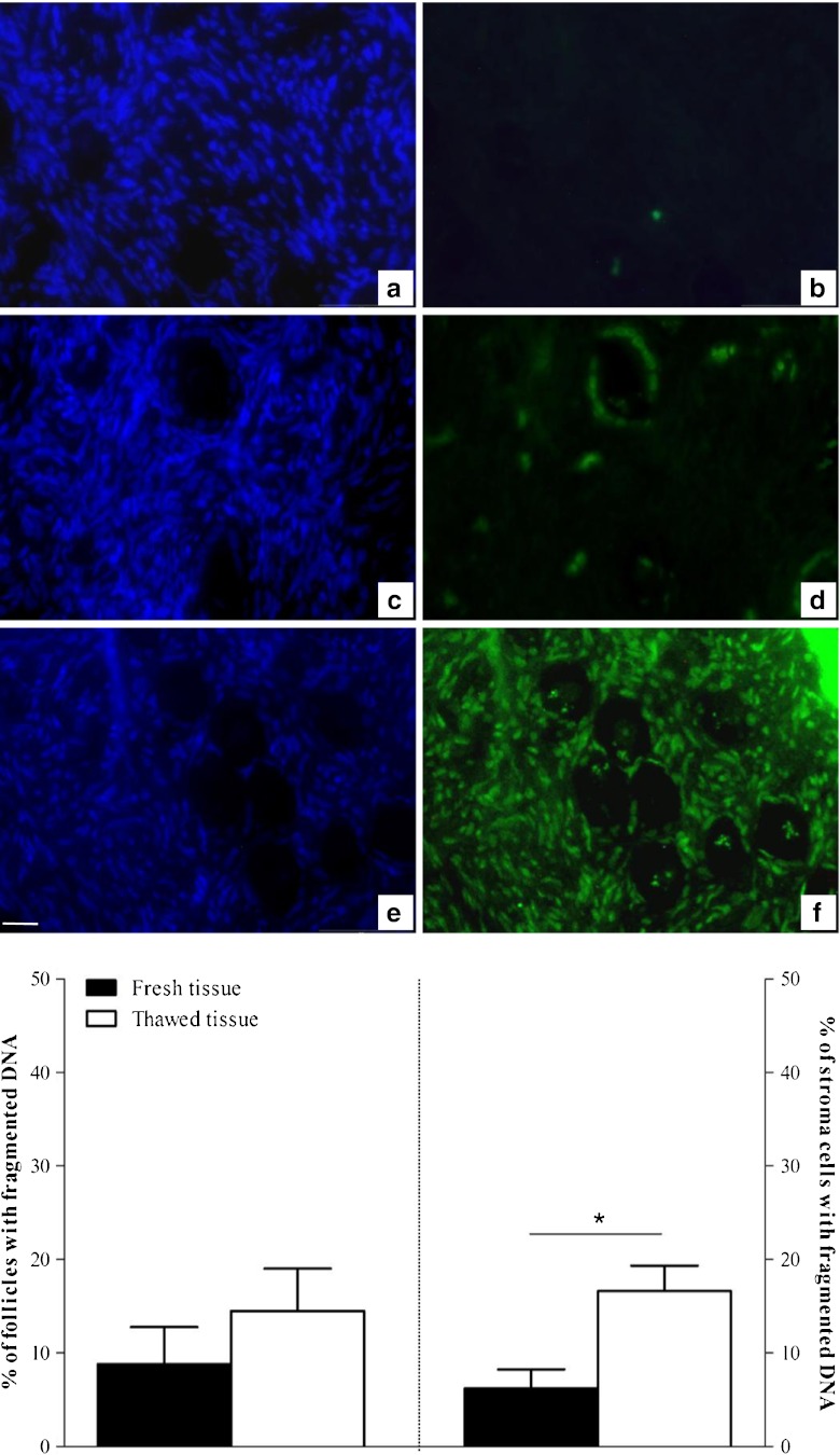
Fig. 1.
DNA fragmentation analysis in follicles and stroma cells. Pictures on the same line are of the same sample but stained using different methods: the Hoechst 33258 method was used to check the location of follicles (left) and the TUNEL method was used to detect DNA fragmentation (right). For each patient, this co-staining was performed on fresh ovarian tissue (a, b) and after slow freezing/thawing using our method (c, d). Cryopreservation induced a slight increase of DNA fragmentation (green fluorescence). e, f DNAse-treated section (TUNEL positive control) both after Hoechst (e) and TUNEL staining (f). Bar = 35 μm. The histograms present the mean percentages (± SEM) of TUNEL-positive follicles (left panel) and stroma cells per high power field (right panel), before (black plots) and after (white plots) cryopreservation (n = 13 patients). *p < 0.05 versus fresh tissue

