Abstract
Purpose
The present study was aimed to find out whether postovulatory aging-induced abortive spontaneous egg activation (SEA) is due to insufficient increase of cytosolic free Ca2+ level.
Methods
Immature female rats (22–24 days old) were subjected to superovulation induction protocol. Eggs were collected 14, 17 and 19 h post-hCG surge to induce in vivo egg aging. The eggs were collected 14 h post-hCG surge and cultured in vitro for 3, 5 and 7 h to induce in vitro egg aging. The morphological changes, rate of abortive SEA, chromosomal status and cytosolic free Ca2+ levels were analyzed.
Results
Postovulatory aging induced morphological features characteristics of abortive SEA in a time-dependent manner in vivo as well as in vitro. The extracellular Ca2+ increased rate of abortive SEA during initial period of culture, while co-addition of a nifedipine (L-type Ca2+ channel blocker) protected against postovulatory aging-induced abortive SEA. However, CI induced morphological features characteristics of egg activation (EA) in a dose-dependent manner. As compare to control, an increase of cytosolic free Ca2+ level (1.42 times) induced abortive SEA, while further increase of cytosolic free Ca2+ level (2.55 times) induced EA.
Conclusion
Our results show that an insufficient cytosolic free Ca2+ level is associated with postovulatory aging -induced abortive SEA, while furthermore increase is required to induce EA in rat.
Keywords: Cytosolic free calcium, Abortive spontaneous egg activation, Postovulatory aging, Rat eggs
Introduction
In most of the mammalian species, ovulated eggs are arrested at metaphase-II (M-II) stage of meiotic cell cycle until fertilization. The fertilizing spermatozoa increases cytosolic free calcium (Ca2+) level and induces egg activation (EA), which is morphologically characterized by an exit from M-II arrest, extrusion of second polar body (PB-II), cortical granules exocytosis and pronuclei formation [1, 2]. The inositol 1,4,5-triphosphate-sensitive stores are the primary source for an increase of cytosolic free Ca2+ level during EA [3–5]. Calcium ionophore A23187 (CI) increases cytosolic free Ca2+ level and induces EA in vitro [6–9].
In the absence of fertilization, postovulatory aging results in abortive spontaneous egg activation (SEA) in several mammalian species including human [2, 10–16]. However, the possible factor(s) that drive postovulatory aging-induced abortive SEA is not yet known. An increase of cytosolic free Ca2+ level is one of the important factors that triggers EA under in vivo as well as in vitro conditions, a possibility exists that, similar to EA, an increase of cytosolic free Ca2+ level may also trigger postovulatory aging-induced abortive SEA. However, there is no evidence to support this possibility. Postovulatory aging-induced abortive SEA deteriorates the egg quality and limits assisted reproductive technologies (ART) such as in vitro fertilization (IVF), intra-cytoplasmic sperm injection (ICSI) and somatic cell nuclear transfer (SCNT) during animal cloning [14, 17–19]. Several drugs such as proteasome inhibitor, MG-135 [20, 21], melatonin [22], verapamil [16] and pyruvate [23] have been used to prevent postovulatory aging-induced abortive SEA in vitro [16, 24].
Rat is an interesting animal model for the study of SEA [14, 16, 25–28], since this process is rapid [29] and abortive [30]. After extrusions of PB-II, rat egg neither form pronuclei and nor proceed to interphase, instead gets arrested at metaphase-III (M-III) like stage [14, 24, 27, 28]. Postovulatory aging is one of the important factors that induce abortive SEA in rat. For example, freshly ovulated eggs recovered after 14 h post human chorionic gonadotropin (hCG) surge are less likely to undergo abortive SEA, while postovulatory aging induces abortive SEA [16, 31–33]. However, factor(s) that drives postovulatory aging-induced abortive SEA are not fully understood. We hypothesize that an insufficient increase of cytosolic free Ca2+ level could be one of the factors associated with postovulatory aging-induced abortive SEA. Hence, present study was designed to investigate the involvement of cytosolic free Ca2+ level during postovulatory aging-induced abortive SEA in rat. For this purpose, postovulatory egg aging was induced in vivo as well as in vitro and morphological changes such as extrusion of PB-II, meiotic status and cytosolic free Ca2+ level were analyzed.
Materials and methods
Unless otherwise stated, all reagents were purchased from Sigma Chemical Co., St. Louis, MO, USA.
Preparation of CI and Fluo-3 AM working solutions
CI was initially dissolved in 100 μL of dimethyl sulfoxide (DMSO) and then in distilled water to get a final concentration of 1 mg/ml (stock solution). The stock solution was further diluted in Ca2+-free medium (Medium-199, AL043A, HiMedia Laboratories, Mumbai, India) to get working concentrations of CI (0.2, 0.3, 0.4 and 0.5 μM). The freshly prepared working concentrations were pre-warmed at 37 °C for 5 min before use. Addition of CI did not alter the osmolarity (290 ± 5 m Osmol) and pH (7.2 ± 0.2) of culture medium. Fluo-3 AM was initially dissolved in 100 μL of DMSO and then in media to get a final concentration of 1 mg/ml (stock solution). The stock solution was further diluted using Ca2+ -free medium to get working concentration (50 μM) of Fluo-3 AM. Since DMSO was used as a solvent for the preparation of CI and Fluo-3 AM stock solutions, an equivalent volume of the highest concentration (0.1 % DMSO) was used in the control group.
Experimental animals
Immature female rats (22–24 days old) of Charles-Faster strain were housed in light-controlled room, with food and water available ad libitum. In order to collect maximum number of ovulated eggs, rats were subjected to superovulation protocol by priming with subcutaneous injection of 15 IU pregnant mare’s serum gonadotropin (PMSG) for 48 h followed by 15 IU hCG for various times depending on the experiments (i.e. 14 h for in vitro studies and 14, 17, and 19 h for in vivo studies). All procedures confirmed to the stipulations of the Institutional Animal Ethical Committee of Banaras Hindu University, Varanasi, UP, India.
Collection and culture of eggs
Ovulated cumulus-enclosed eggs were collected from oviduct using a 26-gauge needle and transferred in pre-warmed Ca2+-free medium. All ovulated cumulus-enclosed eggs were picked up using micropipette (Clay Adams; B&D and Co., NJ) and transferred to fresh culture medium containing 0.01 % hyaluronidase at 37 °C for 3 min for the removal of cumulus cells. The denuded eggs were washed three times with fresh culture medium and then 15–20 denuded eggs were transferred 200 μl of culture medium for in vitro studies. The eggs used for all in vitro experiments were arrested at the M-II stage showing first polar body (PB-I).
Effect of postovulatory aging on morphological changes in egg in vivo
Ovulation occurs in immature rats (22–24 days old) subjected to superovulation induction protocol after 14 h post-hCG surge. In order to induce postovulatory aging, eggs were collected 14, 17 and 19 h post-hCG surge. These eggs were quickly denuded as described above and then observed for their morphological changes using phase-contrast microscope (Nikon, Eclipse; E200, Japan) at 400x magnification.
Effect of extracellular Ca2+ on morphological changes in eggs in vitro
A group of 15–20 eggs (collected 14 h post-hCG surge) was cultured either in Ca2+ -free medium-199 or Ca2+ (1.80 mM) supplemented medium (Medium-199, Cat. no. AL014A; HiMedia Laboratories, Mumbai, India) containing with or without 200 μM nifedipine (a known L-type Ca2+ channel blocker). This concentration of nifedipine has been reported to inhibit SEA in rat eggs cultured in vitro [29]. All cultures were maintained at 37 °C for 3, 5 and 7 h in humidified chamber. At the end of the incubation period, eggs were examined for abortive SEA using a phase-contrast microscope at 400x magnification.
Effect of CI on morphological changes in eggs in vitro
We used CI as a positive control in the present study to compare morphological changes characteristics of EA with postovulatory aging-induced abortive SEA. For this purpose, 15–20 eggs collected after 14 h post-hCG surge were cultured in Ca2+-free medium with or without various concentrations of CI (0.2, 0.3, 0.4 and 0.5 μM) for 3 h in humidified chamber at 37 °C. At the end of the incubation period, eggs were removed, washed with fresh medium and then examined for morphological changes using phase-contrast microscope at 400x magnification.
Analysis of meiotic status of eggs
To analyze the meiotic status, 14–15 eggs (eggs at various stages of meiotic cell cycle) were incubated in medium containing Hoechst stain (0.5 μg/ml of distilled water) for 10 min at 37 °C. At the end of the incubation period, eggs were removed and washed three times with phosphate buffer saline and then examined for chromosomal status under Epi-fluroscence Microscope (Nikon, Eclipse; E-80i, Japan) using Ex350 nm and Em460 nm at 400x magnifications.
Analysis of cytosolic free Ca2+ level using Fluo-3
The cytosolic free Ca2+ level was analyzed in eggs at various stages of meiotic cell cycle following published protocol with some modifications [3, 34, 35]. In brief, eggs (14–15 eggs in each group) were cultured in Ca2+-free medium with or without CI (0.5 μM) and 50 μM Fluo-3 AM for 1 h at 37 °C in humidified chamber. At the end of the incubation period, eggs were removed and washed three times with Ca2+-free medium and observed under Epi-fluroscence Microscope (Nikon, Eclipse; E-80i, Japan) using Ex488 nm and Em525 nm. The corrected total cell fluorescence (CTCF) was calculated following the method published earlier [34] using Image J Software (version 1.44 from National Institute of Health, USA).
Statistical analysis
Data are expressed as mean±standard error of mean (SEM) of triplicate samples. All percentage data were subjected to arcsine square-root transformation before statistical analysis. Data are analyzed either by Student’s t-test or by one-way ANOVA followed by post hoc (Bonferroni) analysis using SPSS software, Version 13 (SPSS, Inc. Chicago, IL). A probability of P < 0.05 was considered as statistically significant.
Results
Postovulatory aging induces abortive SEA in vivo
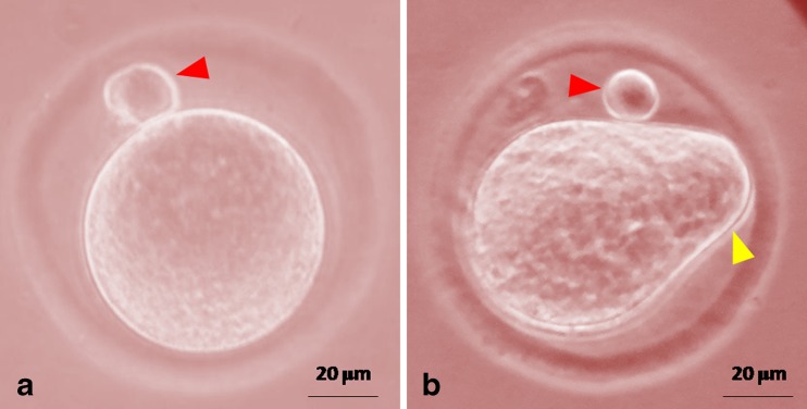
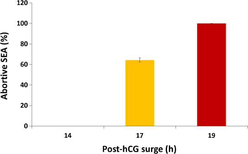
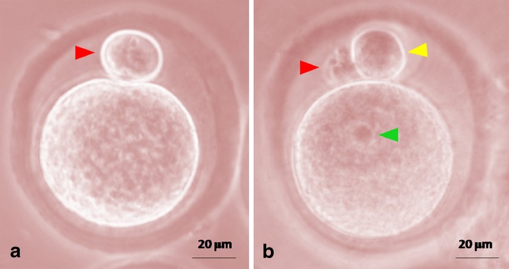
As shown in Fig. 1, newly ovulated eggs collected after 14 h post-hCG surge were arrested at M-II stage of meiotic cell cycle and extruded PB-I (Fig. 1a). Postovulatory aging induced exit from M-II arrest and incomplete extrusion of PB-II, a morphological feature of abortive SEA (Fig. 1b). The rate of abortive SEA was increased in a time-dependent manner if the egg were collected after 17 and 19 h post-hCG surge (One-way ANOVA; F = 1863.4, P < 0.001; Fig. 2). All eggs underwent abortive SEA if collected after 19 h of post-hCG surge.
Fig. 1.
Representative photographs showing postovulatory aging-induced abortive SEA in vivo. a Egg collected 14 h post-hCG surge showing extruded PB-I. b Eggs collected 19 h post-hCG surge showing PB-I (red arrow head) and incomplete extrusion of PB-II (yellow arrow head). Bar =20 μm
Fig. 2.
Postovulatory aging-induced abortive SEA in vivo. Eggs were collected 17 and 19 h post-hCG surge and morphological features of abortive SEA were analyzed. Data are mean (%)±SEM of three replicates and data were analyzed by one-way ANOVA
Extracellular Ca2+ has a role during abortive SEA in vitro
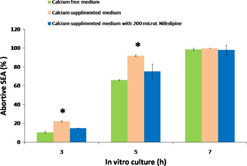
As shown in Fig. 3, postovulatory aging in vitro induced abortive SEA a time-dependent manner in eggs cultured either in Ca2+-deficient medium (one-way ANOVA; F = 4578.5, P < 0.001) or Ca2+ supplemented medium (one-way ANOVA; F = 3154, P < 0.001). Presence of extracellular Ca2+ induced significantly higher rate of abortive SEA during 3 and 5 h of in vitro culture. Almost all eggs underwent abortive SEA after 7 h of in vitro culture in either media suggesting the role of extracellular Ca2+ during postovulatory aging-induced abortive SEA in vitro. However, nifedipine (200 μM) protected extracellular Ca2+ -induced abortive SEA in eggs cultured in Ca2+ supplemented medium (Fig. 3).
Fig. 3.
Postovulatory aging-induced abortive SEA in vitro. Eggs collected 14 h post-hCG surge were cultured in Ca2+-free medium or Ca2+-supplemented medium with or without nifedipine (200 μM) for various times (3, 5 and 7 h). Data are mean (%) ± SEM of three replicates. Data were analyzed by one-way ANOVA followed by post hoc test (Bonferroni). “*” Denotes significant (P < 0.05) increase as compare to Ca2+-free medium
CI induces morphological features characteristics of EA in vitro
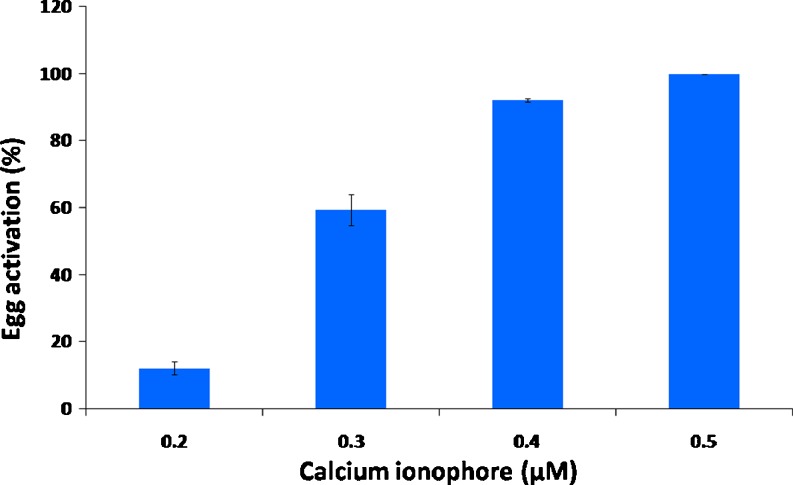
To confirm the role of Ca2+ in inducing morphological features characteristics of EA, we used CI, a known drug that releases Ca2+ from internal stores. As shown in Fig. 4, CI induced morphological features characteristics of EA in a dose-dependent manner (one-way ANOVA; F = 247.23, P < 0.001). Egg collected 14 h post-hCG surge were arrested at M-II stage exhibiting PB-I (Fig. 5a). However, an exit from M-II arrest, complete extrusion of PB-II and pronuclei formation were the morphological features associated with CI-induced EA (Fig. 5b).
Fig. 4.
CI-induced morphological changes characteristics of EA in vitro. Eggs collected 14 h post-hCG surge were cultured in Ca2+ -free medium containing various concentrations of CI for 3 h in vitro. Data are mean (%)±SEM of three replicates and analyzed by one-way ANOVA
Fig. 5.
Representative photographs showing CI-induced morphological changes characteristics of EA in vitro. a Control egg showing PB-I. b CI-induced EA as evidenced by degenerating PB-I (red arrow head), complete extrusion of PB-II (yellow arrow head) and formation of pronuclei (green arrow head). Bar =20 μm
Postovulatory aging induces M-III like arrest
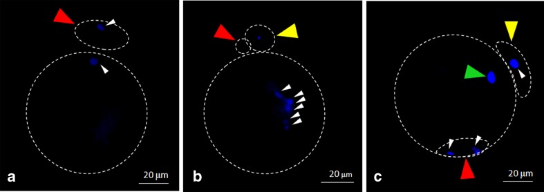
The meiotic status was confirmed during abortive SEA and CI-induced EA using Hoechst stain. As shown in Fig. 6a, control eggs were arrested at M-II stage as evidenced by condensed haploid set of chromosomes in egg cytoplasm as well as in PB-I. Eggs that underwent postovulatory aging–induced abortive SEA showed scattered chromosome in the egg cytoplasm suggesting M-III like arrest and condensed haploid set of chromosomes in PB-II (Fig. 6b). However, eggs underwent CI-induced EA showed pronuclei formation, condensed chromosomes in PB-I and PB-II suggesting the completion of meiosis (Fig. 6c).
Fig. 6.
Representative photographs showing meiotic status of eggs. a Control egg showing condensed chromosome (white arrow heads) in egg cytoplasm as well as in PB-I (red arrow head) characteristics of M-II arrest. b Egg showing scattered chromosomes (white arrow heads) in the cytoplasm suggesting M-III like arrest, extrusion of PB-II (yellow arrow head) and degenerating PB-I (red arrow head). c Egg showing pronuclei formation (green arrow head), complete extrusion of PB-II (yellow arrow head), condensed chromosome (white arrow heads) in PB-I (red arrow head) and PB-II suggesting the completion of meiosis. Bar =20 μm
Postovulatory aging increases cytosolic free Ca2+ level
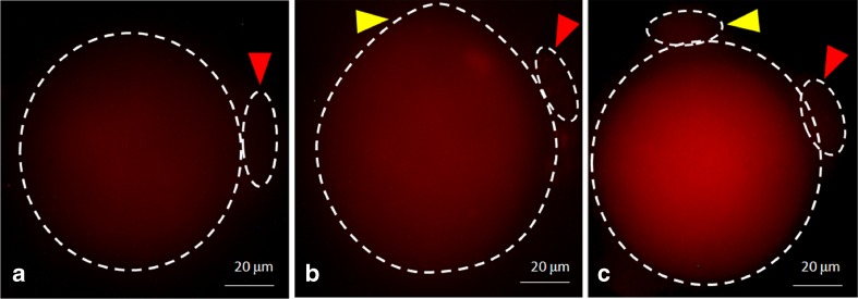
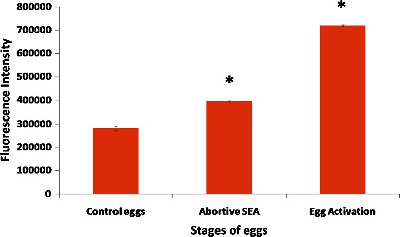
To confirm the role of cytosolic free Ca2+ during postovulatory aging-induced abortive SEA and CI-induced EA, fluorescence intensity of Fluo-3 was analyzed. As shown in Fig. 7, a rise in fluorescence intensity of Fluo-3 was observed in eggs underwent postovulatory aging-induced abortive SEA (Fig. 7b) as compare to control eggs (Fig. 7a). However, further increase of fluorescence intensity was noticed in eggs that underwent CI-induced EA (Fig. 7c). The CTCF analysis using Image J software (Version 1.3) of three independent experiments further confirm that the postovulatory aging significantly (P < 0.001) increased cytosolic free Ca2+ level (1.42 times) and induced abortive SEA, while further increase (2.55 times) was associated with CI-induced EA (Fig. 8).
Fig. 7.
Representative photographs showing fluorescence intensity of Fluo-3 in eggs. a Control egg arrested at M-II stage. b An increase of fluorescence intensity in postovulatory aging-induced abortive SEA. c The fluorescence intensity was further increased in eggs that underwent CI-induced EA. PB-I (red arrow head), PB-II (yellow arrow head). Bar =20 μm
Fig. 8.
The CTCF analysis of fluorescence intensity of Fluo-3 in eggs. Data are mean±SEM of fluorescence intensity using representative eggs of three different experiments. Data were analyzed by Students t-test, “*” Denotes significant (P < 0.001) increase as compare to control eggs
Discussion
Postovulatory aging -induced SEA has been reported in several mammalian species [14, 16, 25, 27, 30]. However, rat is an interesting model because the process of SEA is abortive [30] and aged eggs are arrested at M-III like stage [24]. The freshly ovulated eggs (14 h post-hCG surge) are less likely to undergo abortive SEA as compare to aged eggs in vivo [28, 31, 33]. In the present study, freshly ovulated eggs (collected 14 h post-hCG surge) were arrested at M-II stage and showed extrusion of PB-I. However, postovulatory aging in vivo (egg collected after 17 and 19 h post-hCG surge) as well as in vitro (freshly ovulated eggs collected after 14 h post-hCG surge and their culture under in vitro conditions for 5 and 7 h) induced abortive SEA in a time-dependent manner. These data further support previous observations that the residence of ovulated eggs in the oviduct or culture of freshly ovulated eggs under in vitro conditions induced morphological features associated with abortive SEA [4, 14, 25–27].
Ca2+ plays a major role in the modulation of egg physiology [36]. Data of the present study suggest that the extracellular Ca2+ increased rate of abortive SEA during initial period (3 and 5 h) of in vitro culture, while all eggs underwent abortive SEA if cultured for 7 h either in Ca2+-deficient or Ca2+-supplemented medium. Further, L-type Ca2+ channel blocker such as nifedipine (200 μM) protected against extracellular Ca2+-induced abortive SEA. These data suggest that the L-type Ca2+ channels are still operated in ovulated eggs [37] and nifedipine blocks L-type Ca2+ channels and inhibits abortive SEA in rat eggs [29]. These data together with previous findings suggest that postovulatory aging induces abortive SEA probably by increasing cytosolic free Ca2+ level. The inhibitory effect of nifedipine suggests that it can be used to delay egg aging in vitro during various ART programs.
Intracellular Ca2+ homeostasis is very important in maintaining the physiology of an ovulated egg. An increase of cytosolic free Ca2+ (due to burst from internal stores) induces EA during fertilization [5, 38, 39]. Further, CI increases cytosolic free Ca2+ level [35] and induces EA in several mammalian species [11, 14, 16, 32, 40–42]. In the present study, we used CI as a positive control and data suggest that CI induced morphological features characteristics of EA in a dose-dependent manner. The complete extrusion of PB-II and formation of pronuclei were clearly observed in eggs that underwent CI-induced EA. However, postovulatory aging-induced abortive SEA showed incomplete extrusion of PB-II and did not show pronuclei formation. The chromosomes were scattered in the egg cytoplasm suggesting M-III like arrest in eggs that underwent postovulatory aging-induced abortive SEA. These results together with previous findings suggest that CI induced EA possibly by increasing cytosolic free Ca2+ level in rat egg cultured in vitro. A possibility exists that postovulatory aging-induced abortive SEA might be due to an increase of cytosolic free Ca2+ level. To test this possibility, we analyzed cytosolic free Ca2+ levels and results suggest that the postovulatory aging increased insufficient cytosolic free Ca2+ level (1.42 times as compared to control eggs arrested at M-II stage) and induced abortive SEA, while more increase of cytosolic free Ca2+ level (2.55 times) induced EA. Although the cytosolic free Ca2+ level during postovulatory aging-induced abortive SEA has not been reported in rat eggs so far, an increase of cytosolic free Ca2+ induces EA in mouse eggs [6].
Conclusions
Present study suggests that the postovulatory aging induces insufficient increase of cytosolic free Ca2+ (1.42 times) level leading to abortive SEA, while further increase in its level (2.55 times) induced EA. Further studies are requried to find out the cause(s) for postovulatory aging induced insufficient increase of cytosolic free Ca2+ level in order to protect abortive SEA and to improve ART outcome.
Acknowledgments
The authors are very thankful to Prof. T.G. Shrivastav, Department of Reproductive Biomedicine, National Institute of Health and Family Welfare, Baba Gang Nath Marg, New Delhi-110067, India for providing fluorescence light microscope facility (Nikon, Eclipse; E-80i, Japan).
Footnotes
Capsule
Postovulatory aging induces insufficient increase of cytosolic free Ca2+ level leading to abortive spontaneous egg activation, while further increase of Ca2+ level is required to induce egg activation.
References
- 1.Schultz RM, Kopf GS. Molecular basis of mammalian egg activation. Curr Top Dev Biol. 1995;30:21–62. doi: 10.1016/S0070-2153(08)60563-3. [DOI] [PubMed] [Google Scholar]
- 2.Xu Z, Abbott A, Kopf GS, Schultz RM, Ducibella T. Spontaneous activation of ovulated mouse eggs: time-dependent effects on M-phase exit, cortical granule exocytosis, maternal messenger ribonucleic acid recruitment, and Inositol 1,4,5-trisphosphate sensitivity. Biol Reprod. 1997;57:743–750. doi: 10.1095/biolreprod57.4.743. [DOI] [PubMed] [Google Scholar]
- 3.Carroll J, Swann K. Spontaneous cytosolic calcium oscillations driven by inositol trisphosphate occur during in vitro maturation of mouse oocytes. J Biol Chem. 1992;267:11196–11201. [PubMed] [Google Scholar]
- 4.Miyazaki S, Yuzaki M, Nakada K, Shirakawa H, Nakanishi S, Nakade S, et al. Block of Ca2+ wave and Ca2+ oscillation by antibody to the inositol 1,4,5-trisphosphate receptor in fertilized hamster eggs. Science. 1992;257:251–255. doi: 10.1126/science.1321497. [DOI] [PubMed] [Google Scholar]
- 5.Ciapa B, Chiri S. Egg activation: Upstream of the fertilization calcium signal. Biol Cell. 2000;92:215–233. doi: 10.1016/S0248-4900(00)01065-0. [DOI] [PubMed] [Google Scholar]
- 6.Vincent C, Cheek TR, Johnson MH. Cell cycle progression of parthenogenetically activated mouse oocytes to interphase is dependent on the level of internal calcium. J Cell Sci. 1992;103:389–396. doi: 10.1242/jcs.103.2.389. [DOI] [PubMed] [Google Scholar]
- 7.Liu CT, Chen CH, Cheng SP, Ju JC. Parthenogenesis of rabbit oocytes activated by different stimuli. Anim Reprod Sci. 2002;70:267–276. doi: 10.1016/S0378-4320(01)00185-3. [DOI] [PubMed] [Google Scholar]
- 8.Ito J, Shimada M. Timing of MAP kinase inactivation effects on emission of polar body in porcine oocytes activated by calcium ionophore. Mol Reprod Dev. 2005;70:64–69. doi: 10.1002/mrd.20182. [DOI] [PubMed] [Google Scholar]
- 9.Jilek F, Huttelova R, Petr J, Holubova M, Rozinek J. Activation of pig oocytes using calcium ionophore: effect of protein synthesis inhibitor cyclohexamide. Anim Reprod Sci. 2000;63:101–111. doi: 10.1016/S0378-4320(00)00150-0. [DOI] [PubMed] [Google Scholar]
- 10.Ruddock NT, Machaty Z, Cabot RA, Prather RS. Porcine oocyte activation: roles of calcium and pH. Mol Reprod Dev. 2001;59:227–234. doi: 10.1002/mrd.1027. [DOI] [PubMed] [Google Scholar]
- 11.Ito J, Shimada M, Terada T. Effect of protein kinase C inhibitor on mitogen-activated protein kinase and p34cdc2 kinase activity during parthenogenetic activation of porcine oocytes by calcium ionophore. Biol Reprod. 2003;69:1675–1682. doi: 10.1095/biolreprod.103.018036. [DOI] [PubMed] [Google Scholar]
- 12.Sergeev IN, Norman AV. Calcium as a mediator of apoptosis in bovine oocytes and preimplantation embryos. Endocrine. 2003;22:169–176. doi: 10.1385/ENDO:22:2:169. [DOI] [PubMed] [Google Scholar]
- 13.Ito J, Shimada M, Terada T. Mitogen-activated protein kinase kinase inhibitor suppresses cyclin B1 synthesis and reactivation of p34cdc2 kinase, which improves pronuclear formation rate in matured porcine oocytes activated by Ca2+ ionophore. Biol Reprod. 2004;70:797–04. doi: 10.1095/biolreprod.103.020610. [DOI] [PubMed] [Google Scholar]
- 14.Lu Q, Chen ZJ, Gao X, Ma SY, Li M, Hu JM, et al. Oocyte activation with calcium ionophore A23187 and puromycin on human oocytes that failed to fertilize after intracytoplasmicsperm injection. Zhonghua Fu Chan Ke Za Zhi. 2006;41:182–185. [PubMed] [Google Scholar]
- 15.Ross PJ, Yabuuchi A, Cibelli JB. Oocyte spontaneous activation in different rat strains. Cloning Stem Cells. 2006;8:275–282. doi: 10.1089/clo.2006.8.275. [DOI] [PubMed] [Google Scholar]
- 16.Chaube SK, Dubey PK, Mishra SK, Shrivastav TG. Verapamil reversibly inhibits spontaneous parthenogenetic activation in aged rat eggs cultured in vitro. Cloning Stem Cells. 2007;9:608–617. doi: 10.1089/clo.2007.0001. [DOI] [PubMed] [Google Scholar]
- 17.Fukuda A, Roudebush WE, Thatcher SS. Influences of in vitro oocyte aging on microfertilization in the mouse with reference to zona hardening. J Assist Reprod Genet. 1992;9:378–383. doi: 10.1007/BF01203963. [DOI] [PubMed] [Google Scholar]
- 18.Yanagida K, Yazawa H, Katayose H, Suzuki K, Hoshi K, Sato A. Influence of oocyte preincubation time on fertilization after intracytoplasmic sperm injection. Hum Reprod. 1998;13:2223–2226. doi: 10.1093/humrep/13.8.2223. [DOI] [PubMed] [Google Scholar]
- 19.Hayes E, Galea S, Verkuylen A, Pera M, Morrison J, Lacham-Kaplan O, et al. Nuclear transfer of adult and genetically modified fetal cells of the rat. Physiol Genom. 2001;5:193–04. doi: 10.1152/physiolgenomics.2001.5.4.193. [DOI] [PubMed] [Google Scholar]
- 20.Zhou Q, Renard JP, Friec LG, Brochard V, Beaujean N, Cherifi Y, et al. Generation of fertile cloned rats by regulating oocyte activation. Science. 2003;302:1179. doi: 10.1126/science.1088313. [DOI] [PubMed] [Google Scholar]
- 21.Wu YG, Zhou P, Lan GC, Wang G, Luo MJ, Tan JH. The effects of delayed activation and MG132 treatment on nuclear remodeling and preimplantation development of embryos cloned by electrofusion are correlated with the age of recipient cytoplasts. Cloning Stem Cells. 2007;9:417–431. doi: 10.1089/clo.2006.0023. [DOI] [PubMed] [Google Scholar]
- 22.Tamura H, Takasaki A, Miwa I, Taniguchi K, Maekawa R, Asada H, et al. Oxidative stress impairs oocyte quality and melatonin protects oocytes from free radical damage and improves fertilization rate. J Pineal Res. 2008;44:280–287. doi: 10.1111/j.1600-079X.2007.00524.x. [DOI] [PubMed] [Google Scholar]
- 23.Liu N, Wu YG, Lan GC, Sui HS, Ge L, Wang JZ, et al. Pyruvate prevents aging of mouse oocytes. Reproduction. 2009;138:223–234. doi: 10.1530/REP-09-0122. [DOI] [PubMed] [Google Scholar]
- 24.Galat V, Zhou Y, Taborn G, Garton R, Iannaccone P. Overcoming M-III arrest from spontaneous activation in cultured rat oocytes. Cloning Stem Cells. 2007;9:303–314. doi: 10.1089/clo.2006.0059. [DOI] [PubMed] [Google Scholar]
- 25.Keefer CL, Schuetz AW. Spontaneous activation of ovulated rat oocytes during in vitro culture. J Exp Zool. 1982;224:371–377. doi: 10.1002/jez.1402240310. [DOI] [PubMed] [Google Scholar]
- 26.Kubiak JZ. Mouse oocytes gradually develop the capacity for activation during the metaphase II arrest. Develop Biol. 1989;136:537–545. doi: 10.1016/0012-1606(89)90279-0. [DOI] [PubMed] [Google Scholar]
- 27.Zernika-Goetz M. Spontaneous and induced activation of rat oocytes. Mol Reprod Dev. 1991;28:169–176. doi: 10.1002/mrd.1080280210. [DOI] [PubMed] [Google Scholar]
- 28.Cui W, Zhang J, Lian HY, Wang HL, Miao DQ, Zhang CX, et al. Roles of MAPK and spindle assembly checkpoint in spontaneous activation and M III arrest of rat oocytes. PLoS One. 2012;7:e32044. doi: 10.1371/journal.pone.0032044. [DOI] [PMC free article] [PubMed] [Google Scholar]
- 29.Yoo JC, Smith LC. Extracellular calcium induces activation of Ca2+/calmodulin dependent protein kinase II and mediates spontaneous activation in rat oocytes. Biochem Biophys Res Commu. 2007;359:854–859. doi: 10.1016/j.bbrc.2007.05.181. [DOI] [PubMed] [Google Scholar]
- 30.Chebotareva T, Taylor J, Mullins JJ, Wilmut I. Rat eggs cannot wait: spontaneous exit from meiotic metaphase-II arrest. Mol Reprod Dev. 2011;78:798–07. doi: 10.1002/mrd.21385. [DOI] [PubMed] [Google Scholar]
- 31.Ben-Yosef D, Oron Y, Shalgi R. Low temperature and fertilization-induced Ca2+ changes in rat eggs. Mol Reprod Dev. 1995;42:122–129. doi: 10.1002/mrd.1080420116. [DOI] [PubMed] [Google Scholar]
- 32.Chaube SK, Khatun S, Mishra SK, Srivastav TG. Calcium ionophore-induced egg activation and apoptosis are associated with the generation of intracellular hydrogen peroxide. Free Radic Res. 2008;42:212–220. doi: 10.1080/10715760701868352. [DOI] [PubMed] [Google Scholar]
- 33.Chaube SK, Tripathi A, Khatun S, Mishra SK, Prasad PV, Shrivastav TG. Extracellular calcium protects against verapamil-induced metaphase-II arrest and initiation of apoptosis in aged rat eggs. Cell Biol Int. 2009;33:337–343. doi: 10.1016/j.cellbi.2009.01.001. [DOI] [PubMed] [Google Scholar]
- 34.Gavet O, Pines J. Progressive activation of cyclin B1-cdk1 coordinates entry to mitosis. Develop Cell. 2010;18:533–543. doi: 10.1016/j.devcel.2010.02.013. [DOI] [PMC free article] [PubMed] [Google Scholar]
- 35.Tripathi A, Chaube SK. High cytosolic free calcium level signals apoptosis through mitochondria-caspase mediated pathway in rat eggs cultured in vitro. Apoptosis. 2012;17:439–448. doi: 10.1007/s10495-012-0702-9. [DOI] [PubMed] [Google Scholar]
- 36.Homa ST, Carroll J, Swann K. The role of calcium in mammalian oocyte maturation and eggs activation. Hum Reprod. 1993;8:1274–1281. doi: 10.1093/oxfordjournals.humrep.a138240. [DOI] [PubMed] [Google Scholar]
- 37.Tosti E. Calcium ion currents mediating oocyte maturation events. Reprod Biol Endocrinol. 2006;4:26–34. doi: 10.1186/1477-7827-4-26. [DOI] [PMC free article] [PubMed] [Google Scholar]
- 38.Stricker SA. Comparative biology of calcium signaling during fertilization and egg activation in animals. Develop Biol. 1999;211:157–176. doi: 10.1006/dbio.1999.9340. [DOI] [PubMed] [Google Scholar]
- 39.Tripathi A, Premkumar KV, Chaube SK. Meiotic cell cycle arrest in mammalian oocytes. J Cell Physiol. 2010;223:592–00. doi: 10.1002/jcp.22108. [DOI] [PubMed] [Google Scholar]
- 40.Borges E, PaesdeAlmeidaFerreiraBraga D, CarvalhodeSousaBonetti T, Laconelli A, Jr, Franco JG., Jr Artificial oocyte activation with calcium ionophore A23187 in intracytoplasmic sperm injection cycles using surgically retrieved spermatozoa. Fertil Steril. 2009;92:131–36. doi: 10.1016/j.fertnstert.2008.04.046. [DOI] [PubMed] [Google Scholar]
- 41.Terada Y, Hasegawa H, Takahashi A, Ugajin T, Yaegashi N, Okamura K. Successful pregnancy after oocyte activation by a calcium ionophore for a patient with recurrent intracytoplasmic sperm injection failure, with an assessment of oocyte activation and sperm centrosomal function using bovine eggs. Fertil Steril. 2009;91:e11–e14. doi: 10.1016/j.fertnstert.2008.12.074. [DOI] [PubMed] [Google Scholar]
- 42.Gualtieri R, Mollo V, Barbato V, Fiorentino I, Iaccarino M, Talevi R. Ultrastructure and intracellular calcium response during activation in vitrified and slow-frozen human oocytes. Hum Reprod. 2011;26:2452–2460. doi: 10.1093/humrep/der210. [DOI] [PubMed] [Google Scholar]


