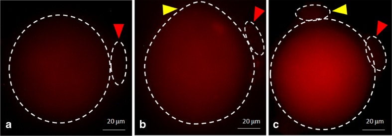
Fig. 7.
Representative photographs showing fluorescence intensity of Fluo-3 in eggs. a Control egg arrested at M-II stage. b An increase of fluorescence intensity in postovulatory aging-induced abortive SEA. c The fluorescence intensity was further increased in eggs that underwent CI-induced EA. PB-I (red arrow head), PB-II (yellow arrow head). Bar =20 μm

