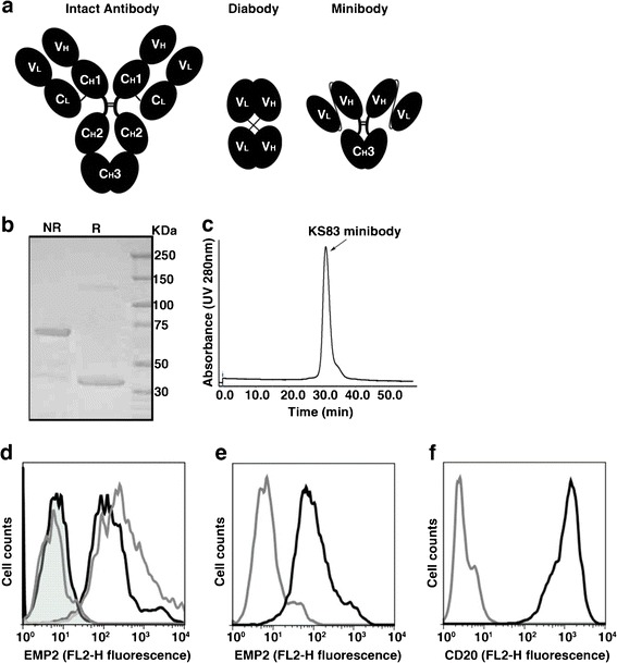
Fig. 2.

Characterization of purified KS83 minibody. a Schematic representation of an intact antibody (150 kDa) and engineered antibody fragments including diabody (scFv dimer, ~55 kDa) and minibody (scFv-hinge-CH3 dimer, ~80 kDa). b Coomassie blue staining after SDS-PAGE of purified KS83 minibody under non-reducing (NR) and reducing conditions (R). Lane 3 Molecular weight marker. c Size-exclusion chromatography of purified KS83 minibody on a Superdex 200 column. Retention time of the sample was compared with appropriate molecular weight standards. d Flow cytometric analysis of HEC1A/EMP2 cells stained with antibody fragments KS83 minibody (black line), KS83 diabody (gray line), or isotype control anti-CD20 minibody (tinted black line) and A10 diabody (tinted gray line) and detected by phycoerythrin-conjugated goat antihuman Fc-specific antibody. e Flow cytometric analysis of murine D2F2 cells stained with KS83 minibody (black line). Anti-CD20 minibody was used as the isotype control (gray line) and detected as above. f Flow cytometric analysis of Ramos cells stained with anti-CD20 minibody (black line). KS83 minibody was used as the isotype control (gray line) and detected as above.
