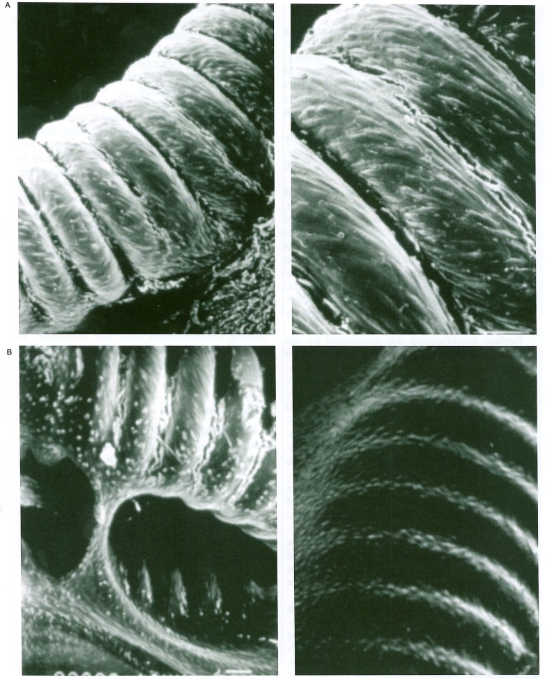
Figure 9.
Scanning electron photomicrographs of the coil coated with type-1 collagen delivered endovascularly into canine carotid artery. A) The coil is completely covered by endothelial cells 2 weeks after delivery (left × 220 and right × 350). B) After 2 months, endothelial cells show a typical mosaic pattern of elongated spindle-shaped cells in flow direction (left × 250 and right × 280).

