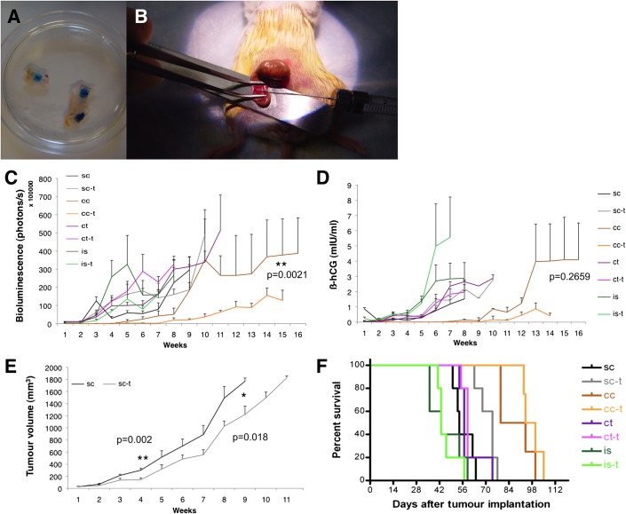
Figure 1.
(A) An injection technique was established by injections of trypan blue into the caecal wall (excised pieces are shown on a petri dish). (B) Exposure of the caecum for orthotopic injections; for successful intra-caecal cell injection, the caecum was placed on a scalpel holder, flattened and stabilised with a forceps. (C) In vivo monitoring: quantification of bioluminescence, (D) ß-human chorionic gonadotropin (ß-hCG) secretion in the urine. (E) subcutaneous tumour sizes. (F) Survival curves. cc, caecal cell implantation; cc-t, caecal cell implantation plus treatment; ct, caecal tissue implantation; ct-t, caecal tissue implantation plus treatment; is, intrasplenic cell implantation; is-t, intrasplenic cell implantation plus treatment.

