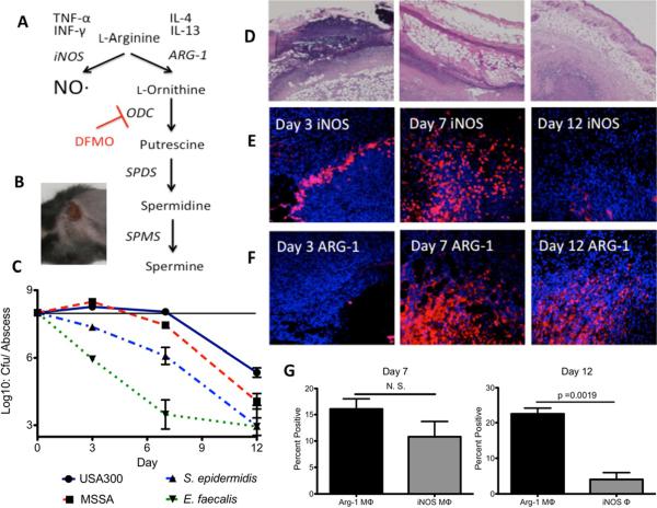
Figure 2. The fate of host arginine during USA300 S. aureus skin infections.
A. Arginine metabolism in response to cytokines during skin infections. B. Dermonecrotic lesion 3 days after subcutaneous inoculation with 1 × 10⁸ cfu of USA300 S. aureus. C. Viable cfu/abscess over time from mice infected with WT USA300 S. aureus, Methicillin-sensitive S. aureus Newman (MSSA), S. epidermidis RP62a or E. faecalis V583. S. epidermidis and E. faecalis abscesses harbored significantly fewer viable cfu compared with USA300 at all three timepoints whereas S. aureus strain Newman was significantly attenuated compared with USA300 at days 7 and 12 (p ≤ 0.05 Mann-Whitney). D. Representative H&E stained tissue imaged at 4× magnification from an infected lesion 3, 7 and 12 dpi. E. Immunofluorescent staining of tissue imaged at 20× from an infected lesion 3, 7 and 12 dpi using α-iNOS antibodies and counterstained with DAPI. F. Similar to Figure 1E except using α-Arg-1 primary antibodies. G. Results of flow cytometery on extracted abscess tissue from day 7 and 12 murine SSTIs. Cells were gated on the living macrophage-like cell population (CD45Hi CD11bHi GR-1Lo-Int CD3Lo B220Lo) and counted for intracellular staining with either anti-Arg-1 or anti-iNOS antibodies.

