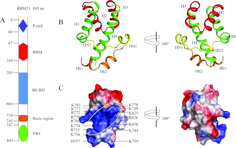
Figure 1. Crystal structure of the RBM25 PWI domain and its flanking basic region.
(A) Schematic diagram of the structural organization of RBM25 and its mutants. RBM25 consists of a proline (P)-rich region and an N-terminal RRM domain, a central RE/RD-rich domain and a C-terminal PWI domain. Amino acid positions are indicated on the left. (B) The structure of the PWI domain and its flanking basic region. The basic region contains two helices (orange). The two views differ by a 180° rotation around the vertical axis. (C) The electrostatic potential surface of the PWI domain and its flanking basic region. The ellipse highlights the surface region rich in positive charge; the black lines mark the basic residues. Single-letter code is used for amino acids. The orientations of (C, left) and (B, left) are the same.

