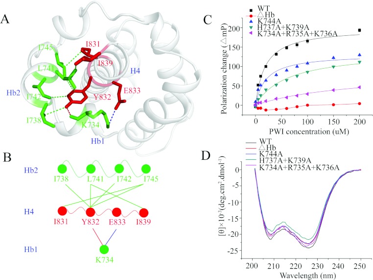
Figure 4. The flanking basic region acts as a co-operative partner with the PWI domain in the binding of nucleic acids.
(A) The interactions between the flanking basic region and helix H4 of the PWI domain. The blue broken line represents the ionic interaction between Glu833 in H4 and Lys734 in Hb1; the red broken line represents the hydrogen bond between Tyr832 in H4 and Lys734 in Hb1; and the green broken lines represent hydrophobic interactions. Single-letter code is used for the amino acids. (B) A brief model of the interactions between the flanking basic region and helix H4 of the PWI domain. The colour code is the same as in (A). (C) The FPAs of the PWI domain and its mutants K744A, H737A+K739A, K734A+R735A+K736A and ΔHb: interactions with 5′ FAM-labelled ssRNA. (D) CD spectra of the PWI domain and its mutants.

