
Figure 3. Kinetic properties of the two major optical neural silencer classes.
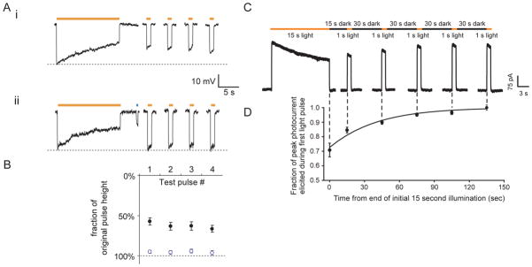
A, (i) Halo-mediated hyperpolarizations in a representative current-clamped hippocampal neuron during 15 seconds of continuous yellow light, followed by four 1-second test pulses of yellow light (one every 30 seconds, starting 10 seconds after the end of the first 15-second period of yellow light). (ii) Halo-mediated hyperpolarizations for the same cell exhibited in (i), but when Halo function is facilitated by a 400-ms pulse of blue light in between the 15-second period of yellow light and the first 1-second test pulse. Adapted from ref. (36). B, Population data for blue-light facilitation of Halo recovery. Plotted are the hyperpolarizations elicited by the four 1-second test pulses of yellow light, normalized to the peak hyperpolarization induced by the original 15-second yellow light pulse (mean + std. err.). Black dots represent experiments when no blue light pulse was delivered (as in Fig. 3Ai). Open blue dots represent experiments when 400 ms of blue light was delivered to facilitate recovery (as in Fig. 3Aii). Adapted from ref. (36). C, Raw current trace of a neuron lentivirally-infected with Arch, illuminated by a 15 s light pulse (575 ± 25 nm, irradiance 7.8 mW/mm2), followed by 1 s test pulses delivered starting 15, 45, 75, 105, and 135 seconds after the end of the 15 s light pulse. Adapted from ref. (42). D, Population data of averaged Arch photocurrents sampled at the times indicated by the vertical dotted lines that extend into Fig. 3C. Adapted from ref. (42).