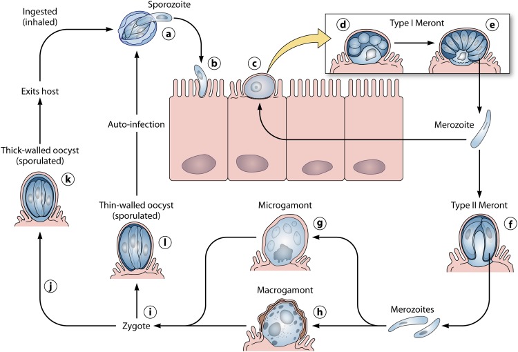
Fig 1.
Schematic representation of the Cryptosporidium parvum life cycle. After excysting from oocysts in the lumen of the intestine (a), sporozoites (b) penetrate host cells and develop into trophozoites (c) within parasitophorous vacuoles confined to the microvillous region of the mucosal epithelium. Trophozoites undergo asexual division (merogony) (d and e) to form merozoites. After being released from type I meronts, the invasive merozoites enter adjacent host cells to form additional type I meronts or to form type II meronts (f). Type II meronts do not recycle but enter host cells to form the sexual stages, microgamonts (g) and macrogamonts (h). Most of the zygotes (i) formed after the fertilization of the microgamont by the microgametes (released from the microgamont) develop into environmentally resistant, thick-walled oocysts (j) that undergo sporogony to form sporulated oocysts (k) containing four sporozoites. Sporulated oocysts released in feces are the environmentally resistant life cycle forms that transmit the infection from one host to another. A smaller percentage of zygotes (approximately 20%) do not form a thick, two-layered oocyst wall; they have only a unit membrane surrounding the four sporozoites. These thin-walled oocysts (l) represent autoinfective life cycle forms that can maintain the parasite in the host without repeated oral exposure to the thick-walled oocysts present in the environment. (Modified from reference 33 with permission.)

