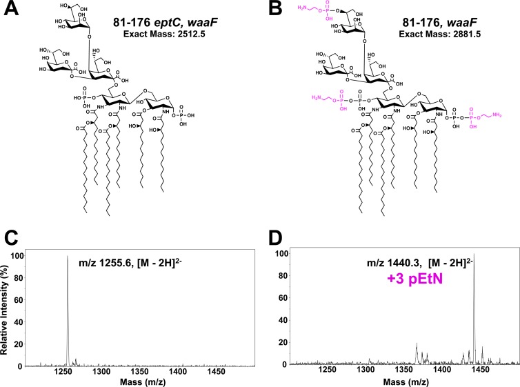
Fig 2.
LC-ESI mass spectra of LOS of C. jejuni strains. (A and B) Proposed structures of the LOS from C. jejuni 81-176 eptC waaF (A) and C. jejuni 81-176 waaF (B). (C and D) LCI-ESI mass spectra of LOS purified from C. jejuni 81-176 eptC waaF (C) and C. jejuni 81-176 waaF (D). The LOS was characterized by LC-ESI–MS in negative mode using collision-induced dissociation as the ion activation method to generate fragmentation ions. Analysis of LOS from strain 81-176 eptC waaF and 81-176 waaF revealed dominant doubly charged molecular ions of 1,255.6 and 1,440.3 [M-2H]2−, respectively. The observed masses are consistent with the proposed structures in panels A and B, and the difference between the dominant molecular ions (184.7 [M-2H]2−) is consistent with the addition of three pEtNs to the LOS in strain 81-176 waaF. The results agree between at least two experimental replicates of a single biological sample.

