Fig 1.

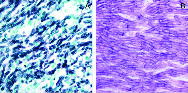
Grocott's methenamine silver (A) or hematoxylin-and-eosin (B) staining of sections from a bronchiectatic cystic structure, showing a mass of interwoven septate hyphae. The hyphae are thin (3 to 5 μm) with parallel walls.

Grocott's methenamine silver (A) or hematoxylin-and-eosin (B) staining of sections from a bronchiectatic cystic structure, showing a mass of interwoven septate hyphae. The hyphae are thin (3 to 5 μm) with parallel walls.