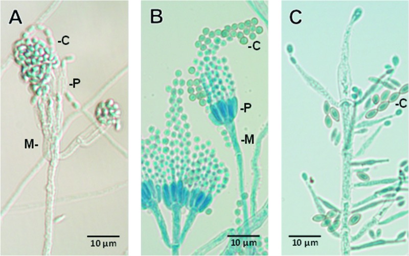
Fig 3.
Photomicrographs show differences in the microscopic morphology of Rasamsonia argillacea (A) from those of a Penicillium species (B) or Paecilomyces variotii (C). All have phialidic conidiogenous cells (P), many of which are supported by metulae (M, cells directly beneath the phialide); however, those in R. argillacea are noticeably roughened. Conidial shape (C) is also distinctive for various species in each of the genera, as shown in the examples above, usually being globose (round) to oval or ellipsoidal in Penicillium or Paecilomyces, respectively, and rectangular to cuneiform (wedge shaped) in R. argillacea. Slide cultures from which photomicrographs were taken were mounted in lactophenol cotton blue.

