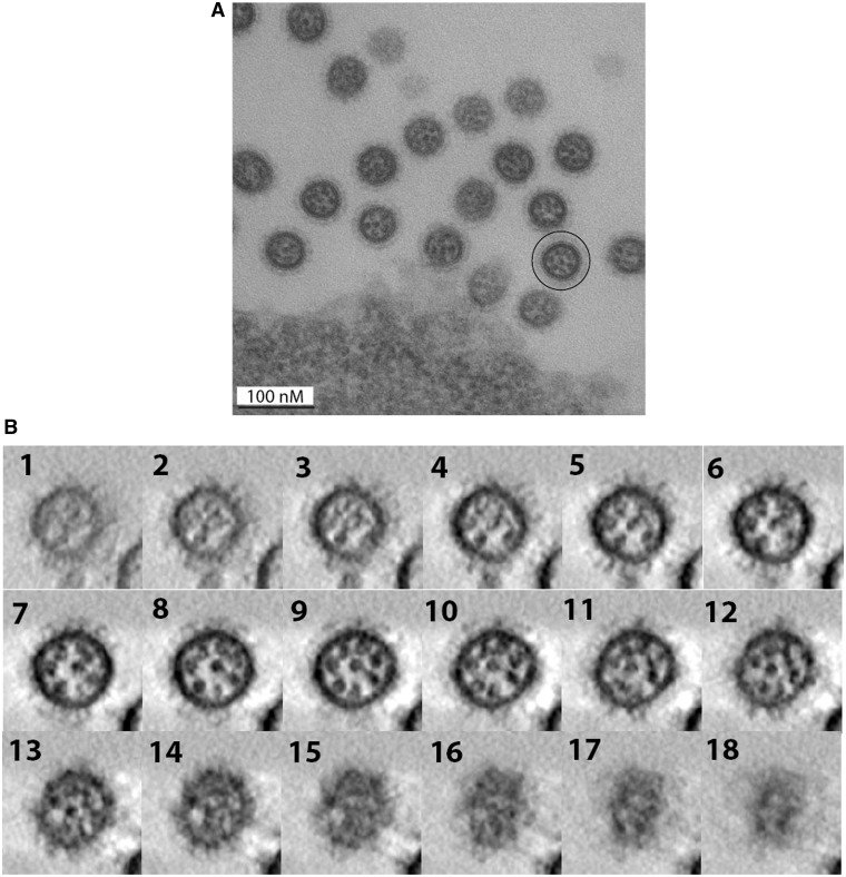
Figure 11.
Electron microscopy (A) and electron tomography (B) of EN viral particles budding from MDCK cells. (A) Cross-sections of budding EN virions. A viral particle in which the eight individual vRNPs form a typical ‘7 + 1’ arrangement is circled. (B) Successive virtual cross-sections of an EN virion, from the bottom to the budding tip of the viral particle.

