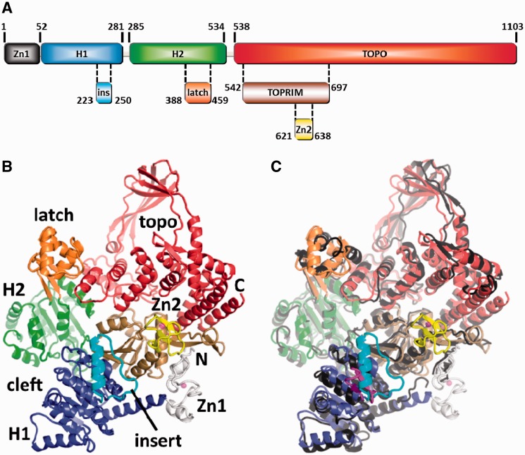
Figure 1.
Overview of reverse gyrase. (A) Domain architecture of reverse gyrase. This color scheme is followed throughout the manuscript. (B) Structure of T. maritima reverse gyrase. The helicase subdomains H1 and H2 are shown in blue and green, respectively. H1 carries an insertion (cyan) that shares similarity with UvrD. The latch region (orange) is inserted in H2. The topoisomerase domain is colored red with the exception of the Toprim subdomain colored in brown. The Toprim domain (from topoisomerase/primase, a doubly wound Rossman fold) serves as a dedicated single-strand DNA-binding site (18). Zinc fingers are drawn in gray (Zn1) and yellow (Zn2). (C) Superposition of the T. maritima and A. fulgidus (PDB-ID 1GKU, dark gray) (11) structures highlights several differences, e.g. the zinc fingers (not resolved in A. fulgidus), the different orientations and folds of the latch and insert domains and a tilt in the topoisomerase domains. The much smaller insert domain of T. maritima reverse gyrase is colored magenta.

