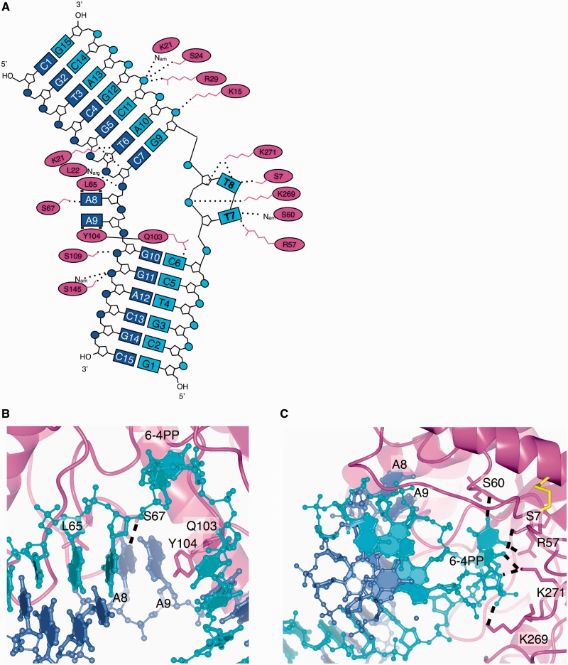
Figure 3.
UVDE-DNA interactions. (A) Schematic representation of UVDE-DNA interactions with the undamaged DNA strand in blue, the damaged strand in cyan and the protein in magenta. The label Nam points out that it is the nitrogen of the amide bond that is involved in hydrogen bonding. In three dimensions, the damaged bases T7 and T8 and the undamaged bases A8 and A9 are actually below the plane of the figure, since they are flipped out of the helix towards one side of the DNA. The residues Q103 and Y104 are together inserting between the two DNA strands near the damage and the two bases opposite to the damage. (B) Detailed view of the pocket for the undamaged bases. Hydrogen bonds are indicated with dashed lines. (C) Detailed view of the pocket for the damaged bases. Hydrogen bonds are indicated with dashed lines.

