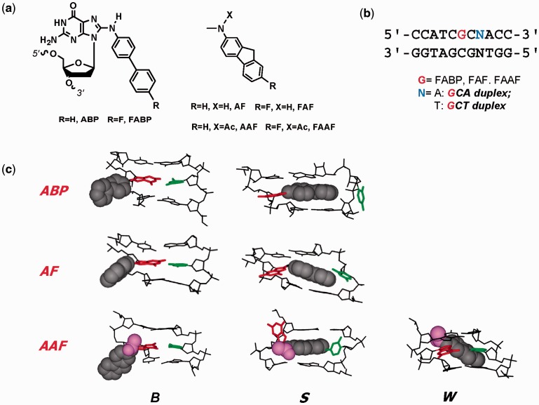
Figure 1.
(a) Structures of ABP [N-(2′-deoxyguanosin-8-yl)-4-aminobiphenyl], AF [N-(2′-deoxyguanosin-8-yl)-2-aminofluorene] and AAF [N-(2′-deoxyguanosin-8-yl)-2-acetylaminofluorene] and their fluoro models, FABP [N-(2′-deoxyguanosin-8-yl)-4-fluoro-4-aminobiphenyl], FAF [N-(2′-deoxyguanosin-8-yl)-7-fluoro-2-aminofluorene] and FAAF [N-(2′-deoxyguanosin-8-yl)-7-fluoro-2-acetylaminofluorene]; (b) 11-mer GCA and GCT duplexes used in this study; (c) Major groove views of the B, S and W-conformers of ABP, AF and AAF. Modified-dG (red), dC (green) opposite the lesion site (orphan C), fluorene (grey CPK), acetyl (AAF only, magenta).

