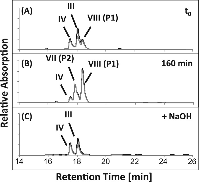
Fig 3.
HPLC chromatograms of a CoA activation assay with cell extracts of Pseudomonas sp. strain Chol1 containing a mixture of Δ1/4- and Δ1,4-3-ketocholate (III and IV in Fig. 1) as the substrates incubated for 0 min (A) and 160 min (B). P1 and P2 represent the CoA esters of Δ1/4- and Δ1,4-3-ketocholate (VII and VIII in Fig. 1), respectively, which are hydrolyzed completely after treatment with NaOH (C). The analysis wavelengths were 245 nm (black) and 260 nm (gray).

