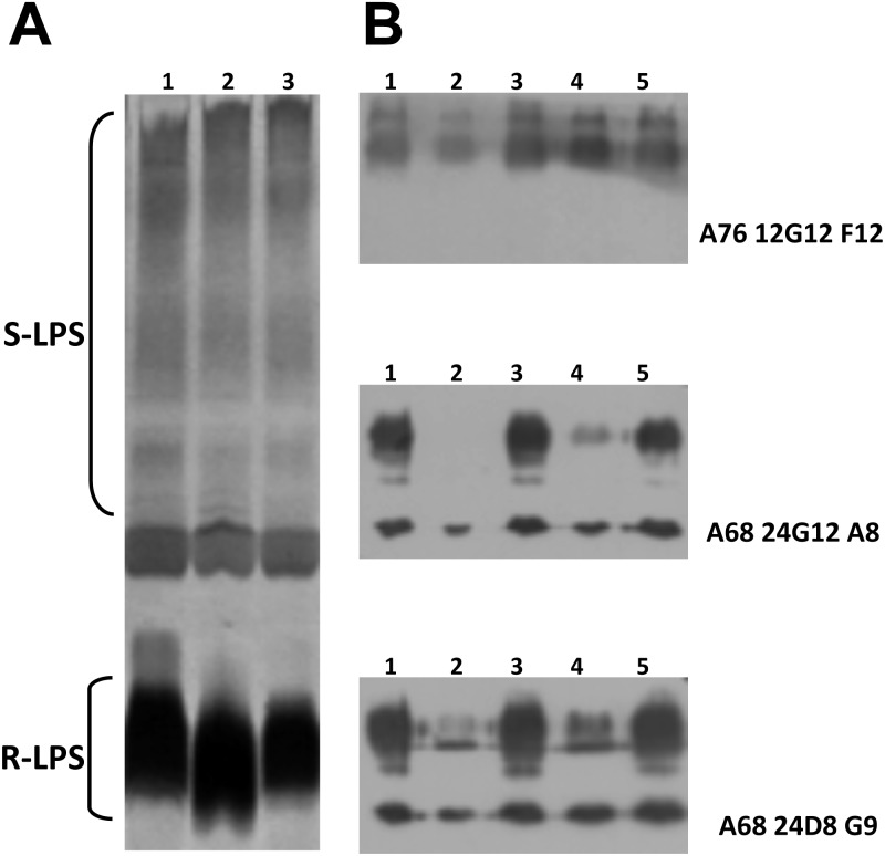
Fig 4.
Modification of LPS in the ΔmucR strain. (A) Silver staining of SDS-proteinase K-treated extracts following electrophoresis on a 16% acrylamide gel. Lane 1, WT; lane 2, ΔmucR; lane 3, ΔmucRKI. (B) Western blot with anti-LPS MAbs. Total extracts were separated on a 15% acrylamide gel by electrophoresis, transferred to nitrocellulose membranes, and probed using anti-O-antigen MAb A76/12G12/F12 and anti-core MAbs A68/24G12/A8 and A68/24D8/G9. Lane 1, WT; lane 2, ΔmucR; lane 3, ΔmucRKI; lane 4, ΔmucR pBBRMCS1; lane 5, ΔmucR pBBRmucR. S-LPS, smooth LPS; R-LPS, rough LPS.

