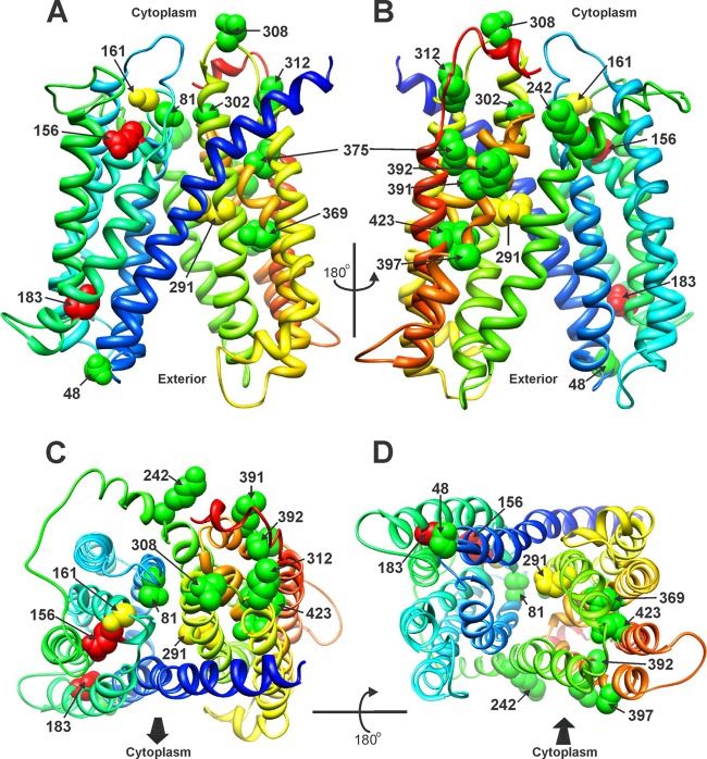
Fig 3.
Residues at which substitution mutations affect efflux function, mapped onto the Phyre2 predicted structure of MepA. Wild-type side chains are depicted. The colors are the same as in Fig. 2, with the exception of residues 48 and 423, which are indicated as fully up function here (green). (A) Front. (B) Rear, rotated 180o from panel A. (C) Top. (D) Bottom. The cytoplasmic face of the protein is toward and away from the reader in panels C and D, respectively, as indicated by the arrows.

