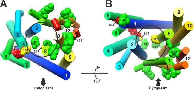
Fig 4.

Phyre2 model of MepA, cylindrical view. Residues and colors are the same as in Fig. 2. (A and B) The cytoplasmic face of the protein is toward (A) and away from (B) the reader, as indicated and correlating with Fig. 3C and D. Helix numbers and specific residues are indicated, as is the presumptive central cavity (oval).
