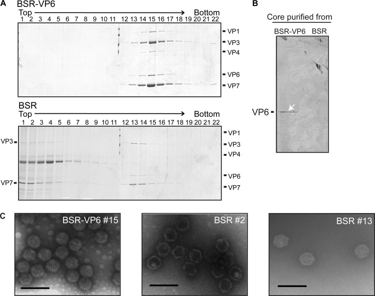
Fig 6.
Analysis of VP6-deficient BTV core particles purified from BSR and BSR-VP6 cells by CsCl gradient centrifugation. (A) Supernatants from BSR-VP6 (top) or BSR (bottom) infected cell lysates were spun down over a 30% (wt/vol) sucrose cushion and subjected to CsCl equilibrium centrifugation. Twenty-two fractions were collected from the top and then analyzed by SDS-PAGE. The gels were stained by Coomassie brilliant blue. (B) BTV core particles purified from BSR-VP6 and BSR cells were resolved by SDS-PAGE, and VP6 was detected by Western blotting using VP6-specific antibody. (C) Electron micrograph of core particles of peak fractions. Bars, 100 nm.

