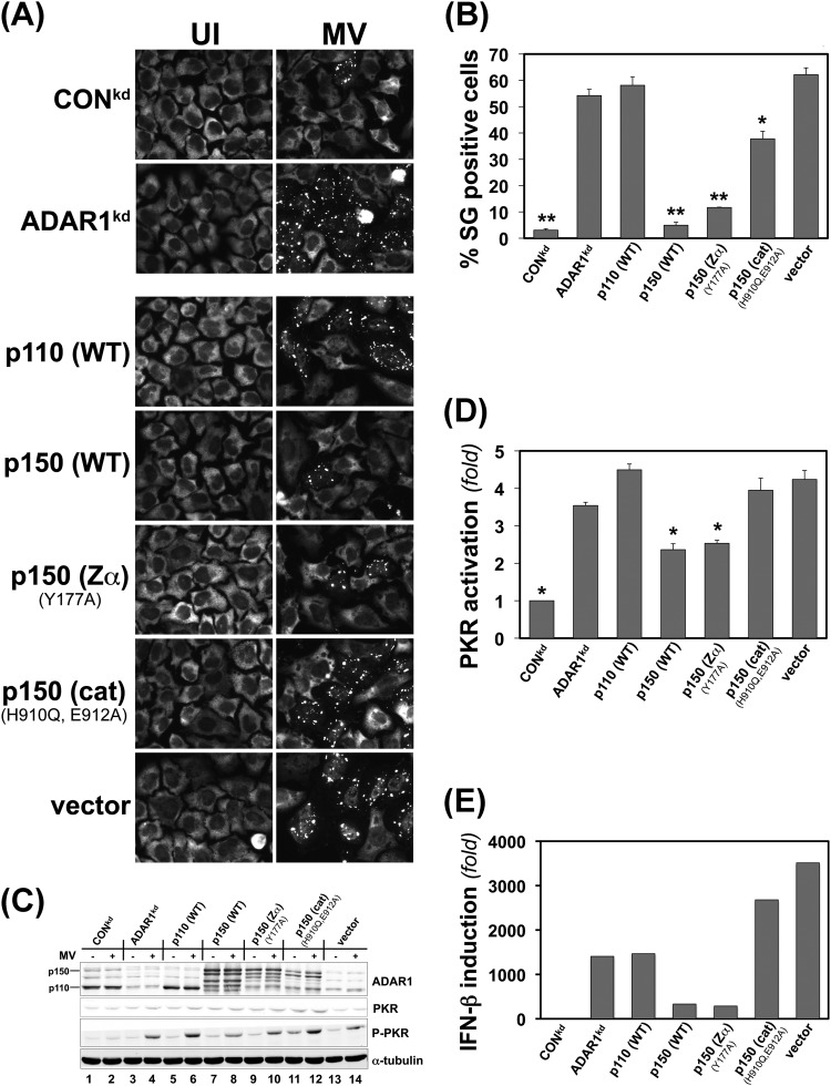
Fig 5.
ADAR1 deaminase activity of the p150 protein isoform is required for suppression of stress granule formation as well as suppression of PKR activation and IFN-β induction following measles virus infection. ADAR1kd cells deficient in p150 and p110 were complemented by transfection with ADAR1 constructs or the vector plasmid as indicated, or ADAR1kd and CONkd cells were also left untransfected as controls. At 24 h after transfection, cells were either infected with WT measles virus (MV) or left uninfected (UI). Infection was with a multiplicity of infection of 1 TICD50/cell for microscopy experiments or 3 TICD50/cell for Western immunoblot assays, IFN-β mRNA quantitation, and yield determinations. ADAR1kd cells complemented with ADAR1 proteins are designated p110 (WT) and p150 (WT) for wild-type protein isoforms and p150 Zα (Y177A) and p150 (cat) (H910Q, E912A) for p150 proteins with mutated domains. Vector, empty vector. (A) At 24 h after infection, cells were analyzed by immunofluorescence microscopy using G3BP1 antibody as a marker for SG formation. (B) Quantitation of SG-positive cells. Wide-field 40× images were obtained, and a minimum of 150 cells were analyzed per experiment for the presence of stress granules as shown in panel A. *, P < 0.001; **, P ≤ 3 × 10⁻⁵ by Student's t test comparing uninfected ADAR1kd cells to infected cells. The results shown are means and standard errors from three independent experiments. (C) Western immunoblot analysis of ectopic ADAR1 expression. At 24 h postinfection, whole-cell extracts were prepared and analyzed using antibodies against ADAR1, PKR, phospho-Thr446-PKR, and α-tubulin as indicated. (D) Quantitation of PKR activation (n-fold) as measured by the level of phospho-Thr446-PKR to total PKR protein determined by Western immunoblot analysis as shown in panel C. *, P < 0.05 by Student's t test to compare the level of P-PKR in infected ADAR1kd cells to infected cells complemented as indicated or to CONkd cells. The results shown are means and standard errors from three independent experiments. (E) IFN-β mRNA levels. At 24 h after infection, total RNA was isolated and IFN-β transcript levels were determined by qPCR and normalized to GAPDH. Results shown are representative of 4 independent experiments.

