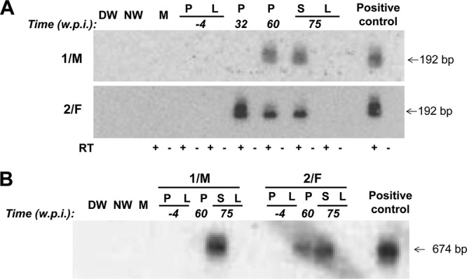
Fig 3.

Detection of WHV RNA and cccDNA in lymphoid cells and tissues (P, PBMC; S, spleen) in the absence of liver (L) engagement in the 1/M and 2/F woodchucks injected with two rounds of multiple low doses of WHV and followed for up to 75 weeks after the first virus injection prior to autopsy. (A) Identification of WHV RNA. DNase-treated total RNA was reversely transcribed (RT+) or not reversely transcribed (RT−) prior to PCR amplification with WHV X gene-specific primers. Positive 192-bp amplicons obtained after nested PCR were identified by NAH. (B) Identification of WHV cccDNA. Total DNA was digested with a single-strand-specific nuclease and amplified by nested PCR with primers spanning the nick region of the WHV genome. NAH was used to confirm the specificity of the 674-bp amplicons and validate controls. Contamination controls shown in panels A and B consisted of water added to direct (DW) and nested (NW) amplification reactions instead of test cDNA or DNA, respectively. Mock (M) samples were extracted and amplified in parallel with test samples. RNA and DNA from PBMC and liver samples collected from the woodchucks prior to first exposure to WHV were used as negative controls, while RNA and DNA from the liver of a serum WHsAg-positive animal with chronic infection were included as a positive control.
