Fig 2.

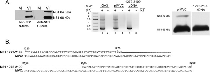
MVC generates two NS1 proteins during viral replication. (A, left) NS1 expression. Immunoblot of mock (M) or virus-infected (VI) WRD cell extracts probed with either an N or C terminus-specific anti-NS1 antibody showing the expression of two distinct NS1 proteins. (Middle) RT-PCR using 5′ and 3′ primers as described in Materials and Methods was done in the presence or absence of reverse transcriptase (RT). cDNA products from the RT-PCR are shown for GA3 virus infection (lane 1), WT pIMVC (lane 2), and the 1272-2199 cDNA (lane 5). (Right) Expression of spliced NS1/pcDNA. Immunoblot of 293T cell extracts transfected with either WT pIMVC or NS1 1272-2199/cDNA probed with an anti-NS1 (C terminus-specific) antibody. (B) NS1 DNA sequence analysis. Alignment of the nucleotide sequences of MVC and NS1 1272-2199/cDNA is shown. The regions depicted are from nt 1220 to 1272 and nt 2168 to 2260. The dashed lines in the NS1 1272-2199/cDNA sequence represent the spliced region from the newly identified 1D′-1A intron.
