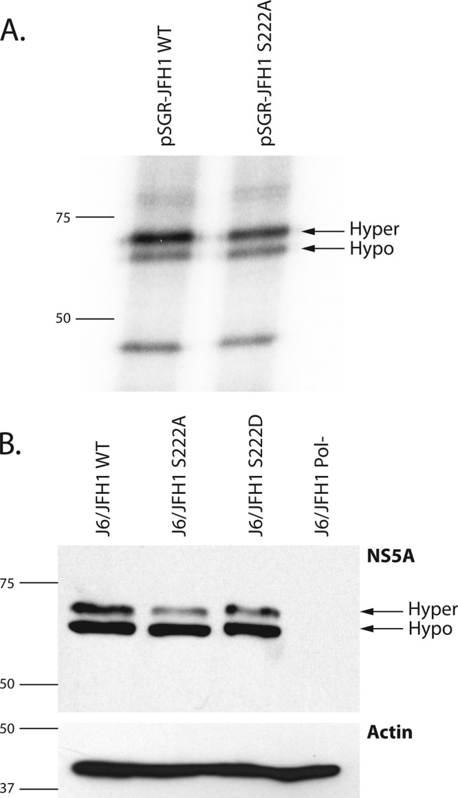
Fig 3.

Metabolic labeling of replicon containing cell lines. (A) Film from 10% SDS-PAGE separation of 32Pi-labeled Huh-7.5 cell lysates from cells harboring wild-type pSGR-JFH1 (pSGR-FJH1 WT) or the pSGR-JFH1 replicon harboring the serine 222-to-alanine mutation (pSGR-JFH1 S222A). Arrows indicate the migration distances of the hypophosphorylated (hypo) and hyperphosphorylated (hyper) forms of NS5A. Numbers and lines to the left of the image indicate the sizes (in kilodaltons) and mobilities of molecular weight markers. (B) Upper panel, Western blot analysis detecting NS5A protein from cells electroporated with HCV J6/JFH1 viral genomic RNA containing S222A, S222D, wild-type, and Pol− RNAs. Arrows indicate the migration distances of the hypophosphorylated (hypo) and hyperphosphorylated (hyper) forms of NS5A. Lower panel, a Western blot of lysates identical to those in the upper panel but probed for the cellular actin protein as a loading control. In both panels, the numbers and lines to the left of the image indicate the sizes (in kilodaltons) and mobilities of molecular weight markers.
