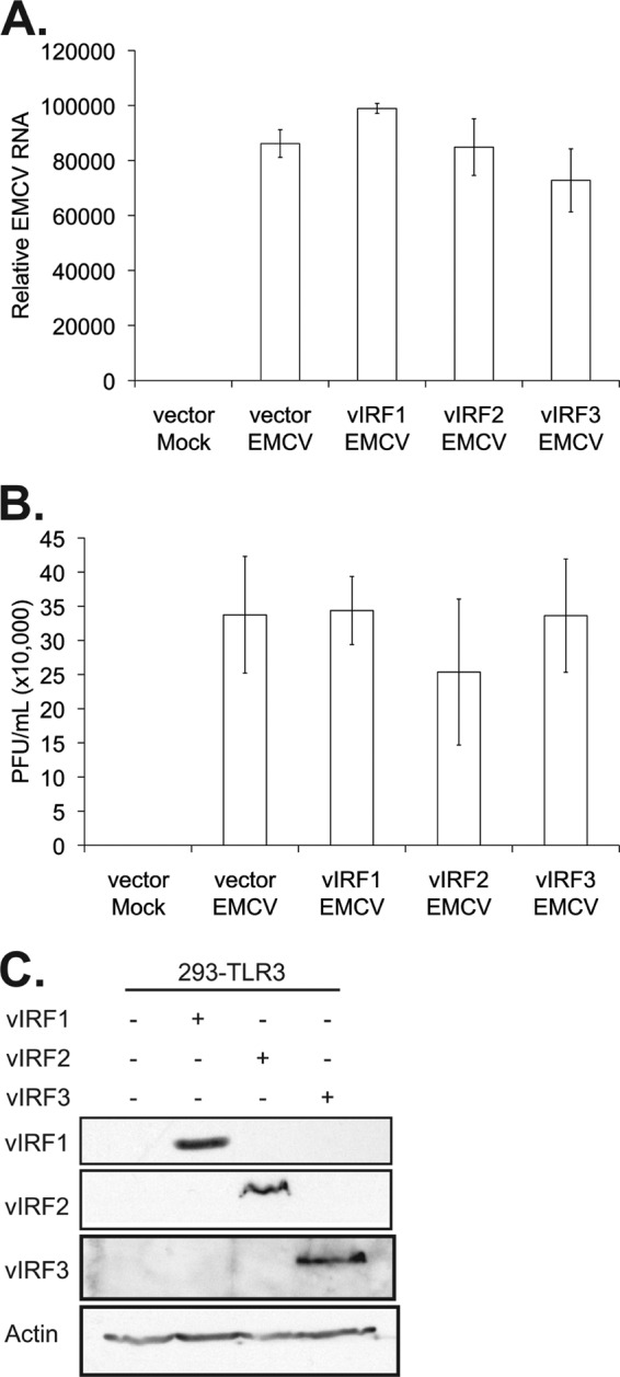
Fig 5.

vIRF expression does not alter EMCV infectivity. 293-TLR3 cells were transfected with the control vector or vectors expressing vIRF1, -2, or -3. Twenty-four hours posttransfection, cells were infected with EMCV at an MOI of 0.001 for 24 h before harvesting. (A) Quantitative RT-PCR was performed on RNA to measure EMCV genetic material, normalized to that of β-actin, represented as the fold increase over vector mock-infected cells. (B) Plaque assays were performed on L929 cells with six different 1:10 serial dilutions of viral supernatant. (C) Lysates harvested at the time of EMCV infection were subjected to SDS-PAGE and immunoblotted with the epitope tags Myc (vIRF1), Xpress (vIRF2), and FLAG (vIRF3) to determine the expression levels of vIRFs posttransfection. Values represent the means plus or minus the standard deviations of the means from triplicate biological replicates.
