Fig 5.

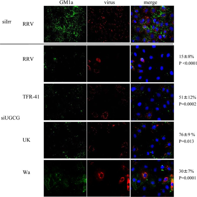
Cells with low ganglioside levels are less susceptible to rotavirus infection. MA104 cells grown in coverslips were transfected with the indicated siRNAs as described in Materials and Methods. Seventy-two hours later, cells were infected with the indicated rotavirus strain (MOI, 3), and eight hours postinfection they were fixed and processed as described in Materials and Methods. Cell membrane ganglioside GM1a was detected with Alexa 488-cholera toxin B subunit (in green); virus-infected cells were detected with polyclonal anti-NSP2 antibody and Alexa 568 anti-rabbit antibody (in red); nuclei were stained with DAPI (in blue). Images were scored independently by two persons, and scores were averaged. In the upper panel a representative staining of cells transfected with siIrr and infected with strain RRV is shown. The lower panel shows representative staining of cells transfected with siUGCG, infected with four different strains. Numbers at the right side correspond to the percentages of infected cells with low levels of ganglioside. Images acquired from at least 3 different experiments were analyzed and counted and represent at least 100 individual cells. Statistical analysis was done using a Student t test, and the values of statistical difference (P) for each virus are shown.
