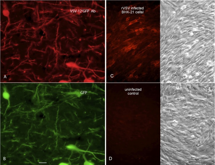
Fig 3.
VSV-12′GFP generates an antibody response against transgene and viral proteins. (A) Antisera were raised against VSV-12′GFP. At a substantial dilution of 1:250,000, the antibody was used to visualize MCH neurons in the brain that expressed avGFP. (B) The GFP expression of the MCH cells immunolabeled in panel A is shown. (C) A VSV that did not express a fluorescent reporter was used to infect BHK cells. The antibody raised against VSV-12′GFP was used to stain infected cells with red immunofluorescence. On the right is the phase image of the same field as shown in the fluorescence micrograph on the left. (D) Control BHK cells show no immunolabeling when stained with the VSV-12′GFP antibody, corroborating the selectivity of the antiserum for the virus protein.

