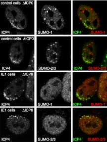
Fig 10.
Inhibition by HCMV IE1 of the recruitment of SUMO-modified species to sites associated with HSV-1 genomes. The upper two panels show the presence of SUMO-1- and SUMO-2-marked proteins in close vicinity of ICP0-null mutant HSV-1 genomes (detected by staining for ICP4) in control cells. The third row shows the absence of local concentrations of SUMO-1 species associated with the ICP4 signal in cells induced to express IE1. The bottom row shows an IE1-expressing cell negative for prominent SUMO-2/3 recruitment. Inductions and infections were performed as noted in the legend to Fig. 9. Samples were stained for ICP4 (MAb 58S) and rabbit antibodies for SUMO-1 or SUMO-2/3 as indicated. Secondary antibodies were anti-mouse Alexa 555 (shown in green) and anti-rabbit Alexa 633 (shown in the red). The quantitative data given in the text were determined by examination of 20 cells in each case.

