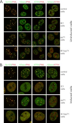
Fig 4.
Immunofluorescence analysis of control, IE1, pp71, and IE1/pp71 cells. (A) Before induction of viral protein expression. (B) After induction with 100 ng/ml doxycycline for 24 h. In both cases, samples were stained as indicated for PML (MAb 5E10) and rabbit antibodies for Sp100, hDaxx, and ATRX, except for the hDaxx-ATRX images, which were stained with rabbit anti-hDaxx and anti-ATRX MAb 39F. Secondary antibodies were anti-mouse Alexa 555 and anti-rabbit Alexa 633 antibodies (shown in the green channel here). Analogous samples were stained for the viral proteins and the ND10 components, indicating the high proportion of cells positive for viral protein expression after induction (see Fig. 1) and the mostly nuclear diffuse distribution of both IE1 and pp71 after induction (images not shown).

