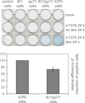
Fig 8.
Derepression of marker gene expression in cells quiescently infected with HSV-1. Cell lines as indicated were either mock infected (upper row) or infected with in1374 at an MOI of 3 for 24 h at the nonpermissive temperature of 38.5°C. The cells in the lowest row were then treated with doxycycline (100 ng/ml) to induce expression of the viral proteins, and all cells were incubated for a further 24 h at 38.5°C prior to staining for β-galactosidase expression. The histogram shows the mean and standard deviation of the proportion of IE1/pp71 cells that were β-galactosidase positive, normalized as a percentage of that in the ICP0 cells after induction. Positive cells were counted in three random views using a 20× objective (about 600 cells total per field of view, of which around 40% of the ICP0-expressing cells were positive).

