
Fig 6.
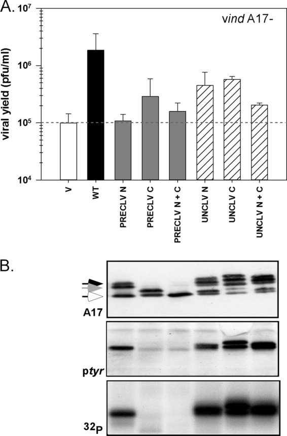
Structure function analysis of the N and C termini of A17. (A) Transient complementation was performed as described previously; cells were infected with vindA17-IPTG and transfected with empty vector (V) or plasmids encoding WT or mutant forms of A17. Viral yield was assessed by a plaque assay; data representing averages of the results of two experiments (with standard errors) are shown. (B) Lysates were prepared from the experiment described for panel A. Samples were analyzed as described for Fig. 5B; the accumulations of total A17 (arrows indicate differentially phosphorylated and/or cleaved species), tyrosine-phosphorylated A17, and phosphorylated A17 were assessed. Immunoblot results for cells transfected with empty vector (V) or WT A17 are shown in Fig. 5B.