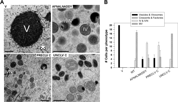
Fig 8.
Structure function analysis of the role of the A17 C terminus in virion morphogenesis. (A and B) BSC-40 cells were infected with vindA17-IPTG and transfected with empty vector or plasmids encoding WT or mutant forms of A17. Cells were harvested at 18 to 24 hpi and processed for conventional electron microscopy. Representative images of the most advanced and most predominant phenotypes are shown in panel A; scale bars represent 0.5 μm. A total of 20 cells were examined and scored as described for Fig. 3C. The scoring categories were vesicles and virosomes, crescents and virosomes, IV and IVN, and MV; data representing averages of the results of two independent experiments (with standard errors) are shown graphically in panel B.

