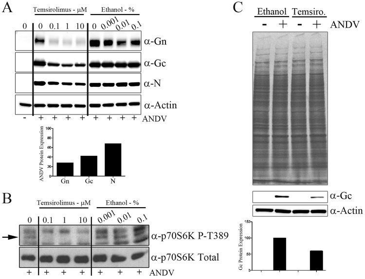
Fig 2.
Temsirolimus reduces ANDV protein levels but not total cellular protein levels. (A) HMVEC-L cells were treated with temsirolimus or ethanol for 1 h, virus was adsorbed in drug-containing medium (MOI of 0.5), and the inoculum was removed and replaced with fresh medium (containing temsirolimus or an equivalent amount of the ethanol vehicle control) after adsorption. At 72 h postinfection, monolayers were lysed and proteins were separated by SDS-PAGE. The expressions of viral nucleocapsid (N) and glycoproteins (Gn and Gc) were observed through Western analysis. The bar graph indicates the densitometric analysis of Gn, Gc, and nucleocapsid protein expressions following treatment with 0.1 μM temsirolimus. Viral protein expression was normalized to actin levels and compared with values for the untreated Gn, Gc, and nucleocapsid protein samples. (B) To confirm the loss of mTOR activity following temsirolimus treatment, HMVEC-L cells were infected with ANDV (MOI of 0.5) and treated with temsirolimus or a carrier. Cells were lysed at 16 h postinfection, proteins were separated by SDS-PAGE, and mTOR activity was assessed through S6K phosphorylation with Western analysis. (C) To measure the effects of temsirolimus on total protein synthesis, HMVEC-L cells were infected as described for panel B. At 8 h postinfection, cells were washed for 30 min with medium lacking cysteine (with or without temsirolimus) and labeled for 16 h with prewarmed cysteine-free RPMI medium containing 100 μCi [35S]cysteine (with or without temsirolimus). Duplicate protein samples were transferred to membranes for Western analysis of viral protein Gc levels. The bar graph indicates densitometric analysis of Gc protein expression normalized to actin levels. Values for control samples treated with ethanol were set at 100%.

