
Fig 4.
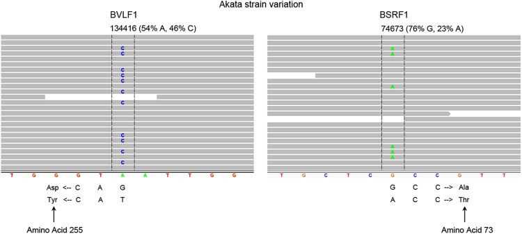
Akata EBV genomes are heterogenous. Screen shots of IGV-displayed aligned reads are shown at the two high-confidence variant locations in the Akata EBV genome. Reads are represented by horizontal gray bars, and variant bases are indicated within the read bars. The genomic position and heterogenous percentage of each variant are indicated above the read bars. The constructed Akata reference genome sequence is displayed at the bottom of the alignments.