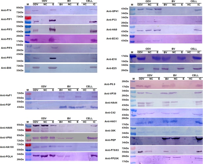
Fig 3.
Western blot analyses of protein localization in the nucleocapsid and envelope fractions of HearNPV BV and ODV. Purified BV and ODV, as well as their nucleocapsid (NC) and envelope (E) fractions, were loaded for Western blot analyses. Healthy cells (HC) and virus-infected cells (IC) were used as negative and positive controls, respectively. The antibodies used for the Western blots are indicated to the left of each blot diagram. M indicates molecular mass markers.

