
Fig 1.
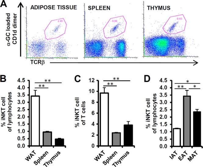
iNKT cells are present in adipose tissue. Eight-week-old C56BL6/J male mice were used for preparation of adipose tissue, spleen, and thymus; n = 5. (A) iNKT cells were detected by staining of α-GC-loaded CD1d dimer and TCR-β. The iNKT cell population is represented as a dot blot graph. (B, C) Percentages of iNKT cells among lymphocytes and T cells in epididymal adipose tissue (EAT), spleen, and thymus. The lymphocyte population was gated by forward scatter (FSC) and side scatter (SSC). (D) Percentages of iNKT cells among lymphocytes in fat depots (inguinal [IAT], epididymal [EAT], and mesenteric [MAT] adipose tissue). *, P < 0.05; **, P < 0.01.