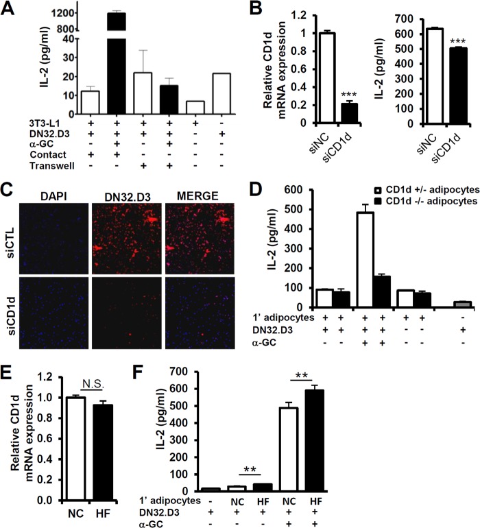
Fig 4.
Adipocyte CD1d regulates iNKT cell activity. (A) DN32.D3 hybridoma cells were used as iNKT cells. Adipocytes and DN32.D3 cells were directly mixed and cocultured in contact or cultured separated by a Transwell membrane, which is cell impermeable with a 0.4-μm pore size. IL-2 secretion by DN32.D3 cells was induced by coculturing with differentiated 3T3-L1 adipocytes which α-GC (100 ng/ml) pretreated for 4 h. (B, left) CD1d expression in negative control (NC) and CD1d siRNA-transfected adipocytes. (Right) IL-2 secretion by DN32.D3 cells upon coculturing with siRNA-transfected 3T3-L1 adipocytes. Adipocytes were pretreated with α-GC (100 ng/ml) for 4 h and, after PBS washing, cocultured with DN32.D3 cells for 6 h. (C) Immunocytochemical analysis of DN32.D3 cells attached to 3T3-L1 adipocytes. Adipocytes were transfected with control siRNA or CD1d siRNA and cocultured with DN32.D3 cells for 24 h. After being washed with PBS, attached red fluorescence-labeled DN32.D3 cells were monitored. (D) The amount of secreted IL-2 was determined by ELISA analysis. Primary adipocytes were isolated from the adipose tissues of CD1d+/− or CD1d−/− mice and cocultured with DN32.D3 cells for 24 h with or without α-GC (100 ng/ml). (E and F) Primary adipocytes were isolated from mice given NCD or HFD for 1 week. (E) Adipocytes derived from epididymal adipose tissues were used. CD1d mRNA level of adipocytes from NCD-fed mice (NC) or from HFD-fed mice (HF). (F) iNKT cells were activated by treatment with α-GC (100 ng/ml)-pretreated adipocytes. Adipocytes and DN32.D3 cells were cocultured for 36 h. n = 5; *, P < 0.05; **, P < 0.01.

