Fig 3.
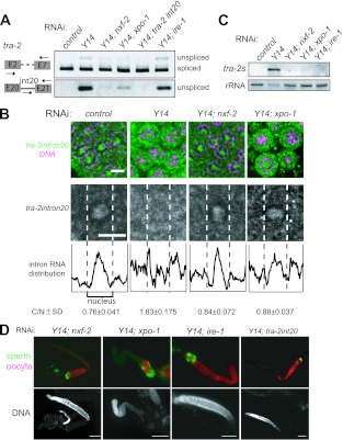
NXF-2 and XPO-1 are responsible for the premature export of unspliced tra-2 RNA. (A) RT-PCR assays were performed to monitor tra-2 RNA expression in the adult hermaphrodites subjected to RNAi as indicated. The primers used for amplification are schematically shown on the left. The amplified products corresponding to the unspliced RNAs were excised and subjected to sequence analysis. Unspliced tra-2 RNA accumulated in Y14(RNAi) animals, which was suppressed by simultaneous RNAi of nxf-2, xpo-1, or tra-2 intron 20 but not ire-1. (B) In situ hybridization of mitotic cells within gonads dissected from adult hermaphrodites subjected to RNAi as indicated. Cells were probed with tra-2 intron 20 (green) followed by DNA staining (magenta), shown as merged views (top). Separate views of single cells probed with tra-2 intron 20 are also shown (middle). The fluorescence intensities in the middle panels were plotted to show the intracellular distribution of the intron (bottom). Dotted lines indicate the borders between the nucleus and cytoplasm. The ratios of the cytoplasmic to nuclear fluorescence intensities for tra-2 intron 20 for five cells were averaged to quantify the intracellular distribution of the intron, shown below (C/N [cytoplasm/nucleus]). Scale bars, 4 μm. (C) Northern blot analysis of tra-2s RNA expression in adult hermaphrodites subjected to RNAi as indicated. 40S rRNA was used as a loading control. (D) Gonad arms were dissected from adult hermaphrodites subjected to RNAi as indicated and stained with the anti-MSP antibody (green) and the anti-RME-2 antibody (red), shown as merged views (top). DAPI-stained views of the same gonad arms are also shown (bottom). Scale bars, 50 μm.

