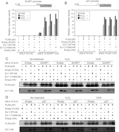
Fig 6.
Raw data from three sets of luciferase assays. H1299 cells were cotransfected with pGL4.12-DUSP1-luciferase (A) or pGL4.12-p21-luciferase (B) and expression vectors for FLAG-p53 and wild-type or C106S DJ-1–HA. Twenty-four hours after transfection, cells were treated with 300 μM H2O2 or with 10 μg/ml cycloheximide (CHX) for 30 min. Luciferase activities were then calculated. (C and D) Proteins were prepared from H1299 cells transfected with FLAG-p53 and wild-type or C106S DJ-1–HA and analyzed by Western blotting.

