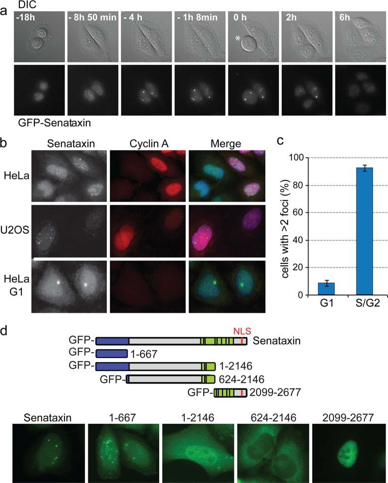
Fig 3.
Immunofluorescence imaging of senataxin foci. (a) Live-cell imaging of HeLa cells transfected with a BAC encoding a FLAP-tagged senataxin. Cells were visualized using differential interference contrast (DIC) and GFP (senataxin) channels. Time zero was defined as the point when the cell indicated with an asterisk entered mitosis. (b) Immunofluorescence imaging of senataxin and cyclin A in fixed HeLa or U2OS cells. Endogenous senataxin was detected using anti-senataxin antibody 1. (c) Quantification of senataxin foci in G1 (cyclin A-negative) and S/G2 (cyclin A-positive) cells (300 cells counted). (d) Identification of the region of senataxin required for S/G2 specific foci. The diagram indicates the five GFP-labeled senataxin constructs expressed in HeLa cells. Senataxin was visualized by GFP immunostaining.

