
Fig 3.
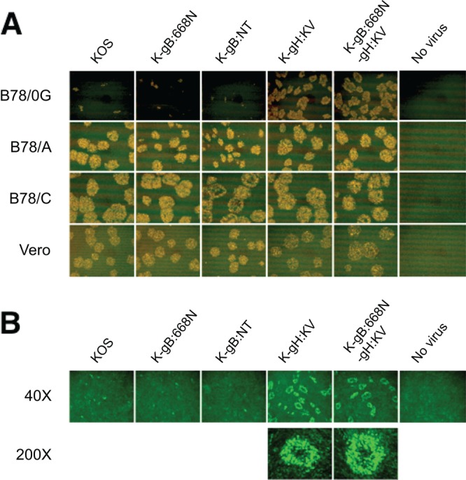
Effects of gB and gH mutations on cell-to-cell spread to and between gD receptor-deficient cells. (A) Vero cells were infected with the viruses indicated above the panels (MOI of 10). Extracellular virus was inactivated by acidic wash, and equal numbers of infected (donor) cells were added onto monolayers of the uninfected cells indicated at the left (acceptor cells). The mixed cultures were overlaid with methylcellulose-containing medium and immunostained for VP16 at 48 hpi. (B) B78/0G cells cultured for 48 h with the donor cells as described for panel A were observed under a fluorescence microscope. Magnifications are indicated at the left. Images are representative of 2 independent experiments.