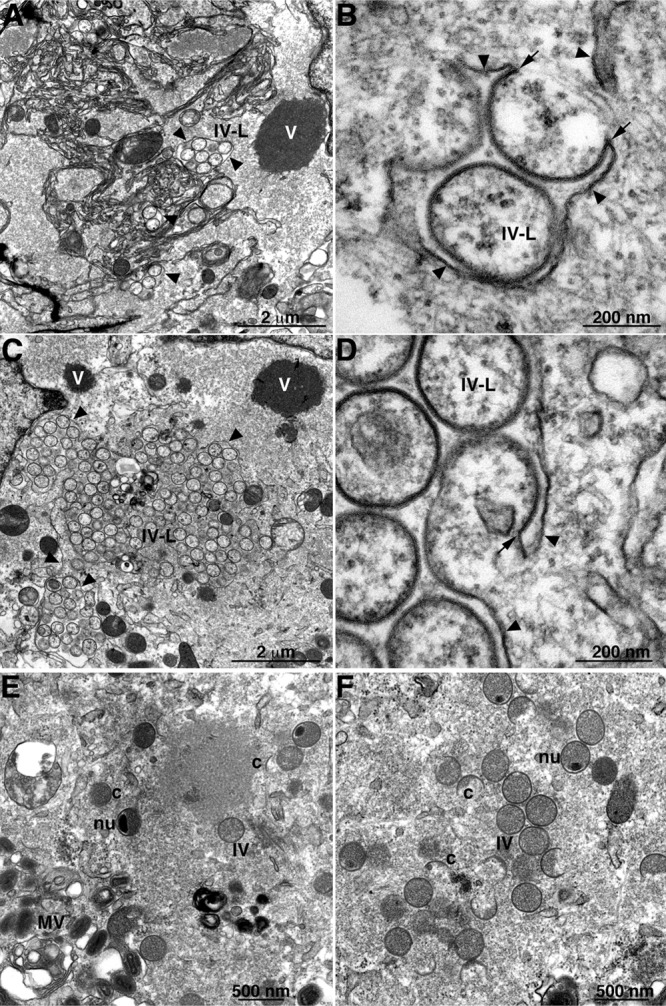
Fig 4.

TEM of cells infected with vΔL2R showing IV-like structures. (A to D) BS-C-1 cells were infected with vΔL2R at a multiplicity of 1 PFU (A and B) and 5 PFU (C and D) per cell. (E and F) Cells were infected with 1 PFU per cell of vΔL2R and transfected 1 h later with a plasmid that expresses L2-HA under the L2R natural promoter. At 20 h after infection, the cells were fixed and prepared for TEM. Abbreviations: c, crescents; IV, immature virion; nu, IV that contains a nucleoid; MV, mature virion; V, dense masses of viroplasm; IV-L, IV-like structures. Arrowheads point to smooth ER membranes; arrows point to the apparent continuity of spicule-coated IV-like structures with smooth ER membranes. The scale bar at the bottom of each panel indicates magnification.
