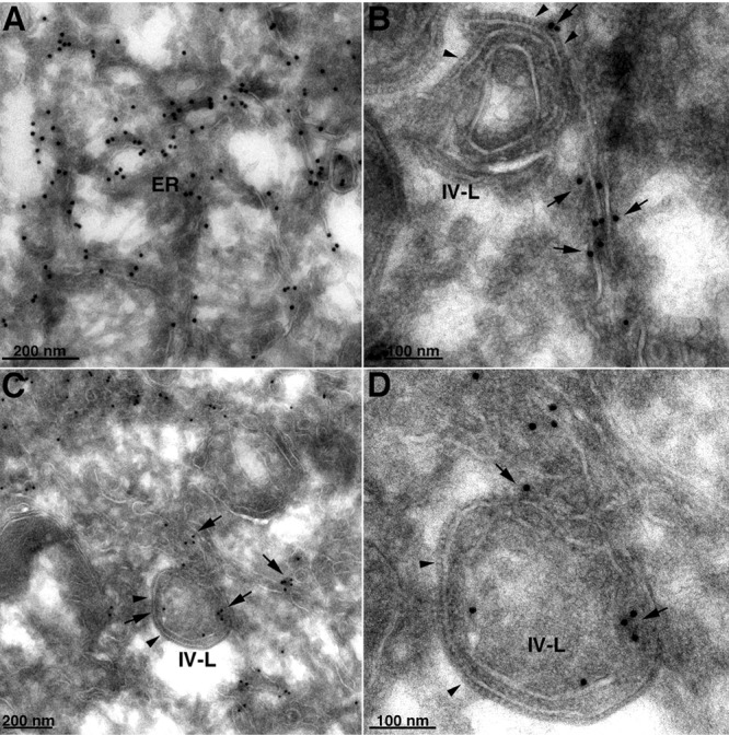
Fig 6.

Localization of calnexin by immunogold TEM in the absence of L2. RK-13 cells stably expressing V5-tagged calnexin were infected with vΔL2R at a multiplicity of 5 PFU per cell. After 21 h, the cells were fixed, cryosectioned, and stained with anti-V5 MAb, followed by rabbit anti-mouse IgG and protein A conjugated to 10-nm gold spheres. (A) Calnexin-staining ER. (B) Image shows an IV-like structure (IV-L). Arrowheads point to the D13 scaffold spicules; arrows point to the adjacent calnexin-staining smooth membranes. (C) Similar to panel B but with calnexin staining of smooth membranes within the IV-like structure. (D) Higher magnification of panel C. The scale bar at the bottom of each panel indicates magnification.
