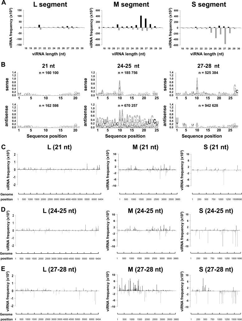
Fig 6.
Production of viRNAs in U4.4 cells infected with RVFV. (A) Size distribution and density of the viRNAs aligning to each segment of RVFV genomic (upper) and antigenomic (lower) polarity in U4.4 cells infected by RVFV ZH548 collected at 48 h p.i.. (B) Logo analysis of 21-, 24/25-, and 27/28-nt viRNA of the genomic (sense) or antigenomic (antisense) polarity. (C to E) Mapping of the 21-nt (C), 24/25-nt (D), or 27/28-nt (E) viRNAs on the RVFV genome.

