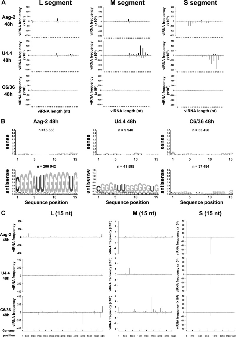
Fig 8.
Analysis of unusually small viRNAs produced in mosquito cells infected with RVFV. (A) Size distribution of the viRNAs of genomic (upper) and antigenomic (lower) polarity in Aag2, U4.4, and C6/36 cells infected by RVFV ZH548, collected at 48 h p.i. (B) Logo analysis of 15-nt viRNA of the genomic (sense) or antigenomic (antisense) polarity. (C) Mapping of the 15-nt-long viRNAs on the RVFV genome in the three cell lines.

