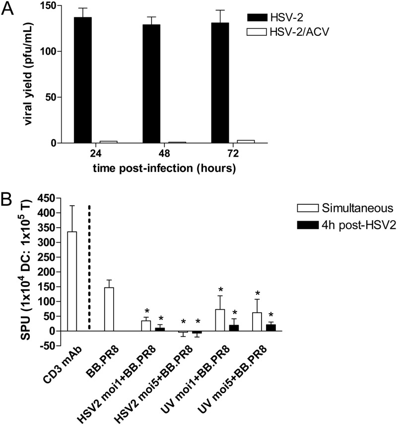
Fig 3.
HSV-2-infected moDCs are impaired in antigen presentation. (A) Immature moDCs or CaSki cells were infected with HSV-2(G) (MOI = 1 PFU/cell) in the absence or presence of acyclovir (ACV) at 100 μg/ml. At the indicated times p.i., culture supernatants were collected, and viral yields were quantified by performing plaque assays on Vero cells. The results are presented as PFU/ml and are means + the SD obtained from five independent experiments conducted in duplicate. (B) 104 human moDCs were exposed to influenza virus BB.PR8 and HSV-2(G) simultaneously or to influenza virus BB.PR8 at 4 h post-HSV-2(G) challenge and then cultured with autologous CD14− T cells at a 1:10 DC/T cell ratio in the presence of ACV (200 μg/ml). IFN-γ release was measured at 40 h postcoculture. T cells stimulated with a CD3 MAb served as a positive control for the ELISpot assay (far left). The results are presented as means + the SEM spot-forming units (SPU) obtained in five independent experiments, each performed in triplicate. The asterisks indicate significant differences relative to influenza virus-challenged DCs (P < 0.05).

