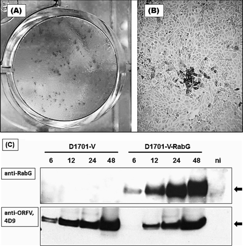
Fig 1.
Expression of RABV G protein in D1701-V-RabG-infected Vero cells. Panels A and B demonstrate IPMA staining of recombinant ORFV plaques expressing RABV glycoprotein. Two days after infection, IPMA shows dark (brownish)-stained positive virus plaques expressing RabG (A). Cells expressing RabG can be easily discriminated from negative cells (panel B, 40-fold microscopic magnification). (C) Western blot analysis to detect the expressed RabG. Protein lysates were prepared at the indicated time points after infection with D1701-V as negative controls, with D1701-V-RabG (MOI of 1.0), or from noninfected cells (ni). The RabG protein 62 kDa in size (arrow) was specifically detected with the polyclonal antiserum G154-3 (1:10,000 diluted).

